ngal light-excited chemiluminescence detection kit, its preparation and use method

A chemiluminescence detection and light excitation technology is applied in the field of lipocalin content detection, which can solve the problems of easy environmental interference and low sensitivity, and achieve the effects of improved detection specificity, short coupling time and high reaction efficiency.

Active Publication Date: 2016-05-18
WUHAN LIFE ORIGIN BIOTECH LTD
View PDF6 Cites 0 Cited by
  • Summary
  • Abstract
  • Description
  • Claims
  • Application Information

AI Technical Summary

Problems solved by technology

[0004] In order to solve the above technical problems, the present invention provides a NGAL light-excited chemiluminescence detection kit, its preparation and use method, which solves the problems of low sensitivity of the detection method in the prior art and the detection is easily disturbed by the environment

Method used


Image

  • ngal light-excited chemiluminescence detection kit, its preparation and use method
  • ngal light-excited chemiluminescence detection kit, its preparation and use method
  • ngal light-excited chemiluminescence detection kit, its preparation and use method

Examples


Embodiment 1

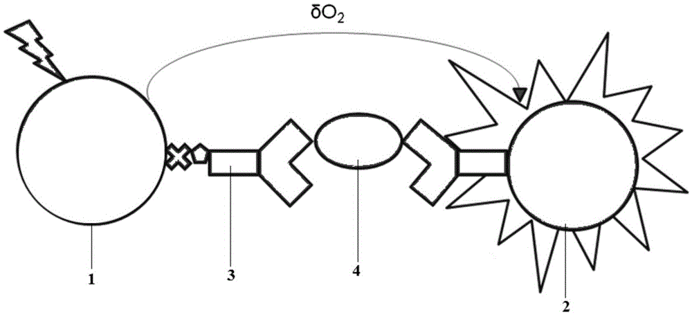
[0028] 1. Luminescent microspheres are connected to antibodies:

[0029] (1) Preparation of antibody and microspheres: Add 0.1 mg of antibody to an ultrafiltration tube, centrifuge for 8 minutes, wash with buffer (pH 8.0) PBS buffer for 6 times, and dilute the antibody to 1 mg / ml for use. The microspheres were resuspended in PBS (pH 8.0) at 20 mg / ml.

[0030] (2) Activate the luminescent microspheres (return all reagents to room temperature before use)

[0031] Measure 1mg of luminescent microspheres, add 1mL of PBS (pH7.4) and vortex, add the suspension into a 1.5mL centrifuge tube, centrifuge at 12000g, carefully suck out and discard the supernatant. Add 100 μL of washing buffer PBS (pH7.4), 0.05% Tween-20 to suspend, shake and sonicate, centrifuge at 12000g, carefully suck out and discard the supernatant. Add 1 mL of PBS buffer (pH6.2), then add 10 μL of freshly prepared EDC (50 mg / mL), then add 10 μL of freshly prepared 50 mg / mL Sulfo-NHS, shake at high speed for 30 seco...

Embodiment 2

[0041] 1. Luminescent microspheres are connected to antibodies:

[0042] (1) Preparation of antibody and microspheres: Add 0.1 mg of antibody to an ultrafiltration tube, centrifuge for 8 minutes, wash with buffer (pH 8.0) PBS buffer for 6 times, and dilute the antibody to 1 mg / ml for use. The microspheres were resuspended in PBS (pH 8.0) at 20 mg / ml.

[0043] (2) Activate the luminescent microspheres (return all reagents to room temperature before use)

[0044] Measure 1mg of luminescent microspheres, add 1mL of PBS (pH7.4) and vortex, add the suspension into a 1.5mL centrifuge tube, centrifuge at 12000g, carefully suck out and discard the supernatant. Add 100 μL of washing buffer PBS (pH7.4), 0.05% Tween-20 to suspend, shake and sonicate, centrifuge at 12000g, carefully suck out and discard the supernatant. Add 1 mL of PBS buffer (pH6.2), then add 10 μL of freshly prepared EDC (50 mg / mL), then add 10 μL of freshly prepared 50 mg / mL Sulfo-NHS, shake at high speed for 30 seco...

Embodiment 3

[0054] Kit preparation

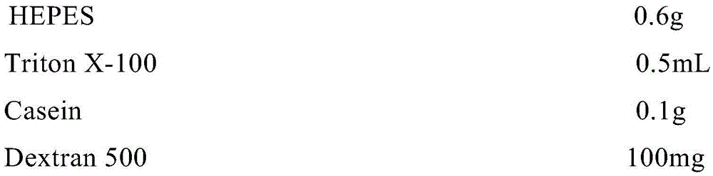
[0055] Take each component according to the stated dosage:

[0056]

[0057] Add 90mL of water to dissolve, adjust the pH to 7.4, make up to 100mL with water, and make the analysis buffer.

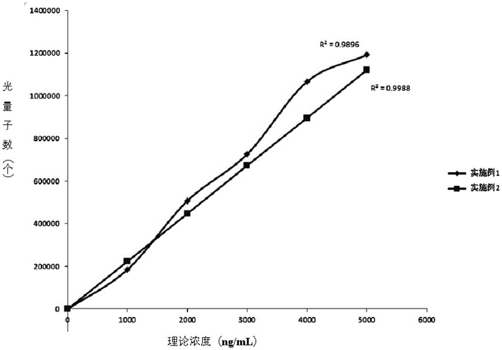
[0058] (1) Dilute the concentration of the luminescent microspheres (prepared in Example 1 or 2) coupled with the NGAL antibody to 50 μg / mL with an analysis buffer;

[0059] (2) Use the analysis buffer to dilute the biotinylated antibody, and adjust the antibody concentration to 0.5mg / mL;

[0060] (3) The assay buffer was used to dilute the coupled avidin-labeled photosensitive microspheres to 80 μg / mL.


PUM

PropertyMeasurementUnit
Sensitivityaaaaaaaaaa

Abstract

The invention relates to a method for preparing an NGAL (Neutrophil Gelatinase Associated Lipocalin) optical excitation chemiluminescence detection kit. The method comprises the following steps: activating a luminous microsphere, namely activating the luminous microsphere through prepared carbodiimide and Sulfo-NHS solutions in a PBS (Phosphate Buffer Saline) buffer solution, wherein the luminous microsphere is a carboxyl modified luminous microsphere, and the luminous microsphere is coated with dimethylthiophene, anthracene and rubrene and carried with an europium chelate; coupling the luminous microsphere with an anti-NGAL antibody, namely selecting the carboxyl modified luminous microsphere, carrying out mixing reaction on the anti-NGAL monoclonal antibody and the activated carboxyl modified luminous microsphere to couple the anti-NGAL antibody to the luminous microsphere, and adding freshly prepared carbodiimide at the same time while mixing the anti-NGAL antibody and the activated luminous microsphere; labeling the anti-NGAL antibody through biotin; and labeling a photosensitive microsphere through avidin. The invention also relates to preparation and use methods of the NGAL optical excitation chemiluminescence detection kit. The methods disclosed by the invention solve the problems that the sensitivity is low and the detection is easily interfered by the environment in a detection method in the prior art.

Description

technical field [0001] The invention relates to the field of detection of lipocalin content, in particular to a light-excited chemiluminescence detection kit for neutrophil gelatinase-associated lipocalin (NGAL) in human body, and its preparation and use methods. Background technique [0002] Currently the most commonly used detection methods for NGAL are colloidal gold immunoassay and chemiluminescence immunoassay (CLIA); [0003] But the applicant finds that these methods have some defects when using colloidal gold and CLIA methods: the detection sensitivity of colloidal gold is not high, and cannot be accurately quantified; there are many influencing factors of CLIA, such as being susceptible to environmental interference, short luminescence time, and being Non-open reagents, high reagent prices. Contents of the invention [0004] In order to solve the above technical problems, the present invention provides a NGAL light-excited chemiluminescence detection kit, its pre...

Claims


Application Information

Patent Timeline
Patent Type & Authority Patents(China)
IPC IPC(8): G01N33/68
CPCG01N33/532G01N33/54393G01N33/92
Inventor 华权高来祥兵徐春雷沈鹤霄许可
Owner WUHAN LIFE ORIGIN BIOTECH LTD
Why Eureka
Try Eureka