Application of human derived HSF2 as specific diagnosis molecular marker of ulcerative colitis
A technology of ulcerative colitis and molecular markers, applied in the field of biomedicine, can solve problems such as no reports and no specific diagnostic markers for UC.
- Summary
- Abstract
- Description
- Claims
- Application Information
AI Technical Summary
Problems solved by technology
Method used
Image
Examples
Embodiment 1
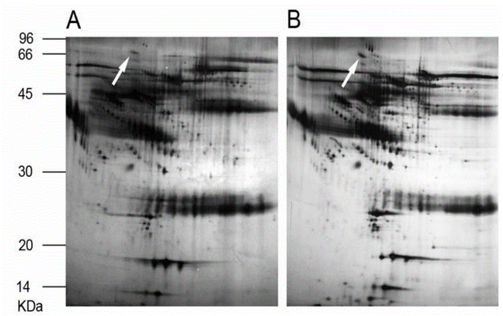
[0013] Example 1: Difference analysis of protein expression in serum of patients with ulcerative colitis (UC) and healthy volunteers.
[0014] 1. Collection and processing of serum samples
[0015] Take 5ml of fasting venous blood from the research subjects, place it at room temperature for 30min, centrifuge at 2000rpm for 20min, and collect serum (cannot be hemolyzed). Serum samples from 40 cases in the same group were mixed uniformly with 30 ul from each case. Albumin and IgG were then removed from serum samples using an Albumin / IgG Removal Kit. The sample protein was then concentrated using a protein precipitation kit.
[0016] 2. The first solid-phase gradient isoelectric focusing electrophoresis and the balance of the gel strip:
[0017] (1) Take a small tube (1ml / tube) of Hydration Loading Buffer (I) (no DTT, no Bio-Lyte) stored at -20°C from the refrigerator, and dissolve at room temperature. Add 0.01g DTT and 25μl Bio-Lyte to the small tube, and mix thoroughly. Ta...
Embodiment 2
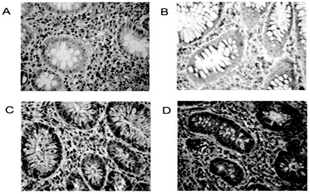
[0051] Example 2: Immunohistochemical detection and analysis of differential expression of HSF2 in mucosal tissue of patients with different degrees of UC. 1. Slicing
[0052] All biopsy tissues were thawed after being taken out from the -80°C refrigerator, embedded with frozen section embedding medium, frozen in liquid nitrogen, and quickly sectioned on a constant temperature cryostat at -20°C. A tissue slice, pasted on a detachment-proof glass slide, fixed in acetone at 4°C for 15 minutes, then taken out, and quickly placed in a -20°C refrigerator for storage after the acetone was completely evaporated.
[0053] 2. Immunohistochemical experimental steps
[0054] (1) Place the slides in a 60°C oven overnight. The next day, the slices were taken out and placed in xylene liquid I and II for deparaffinization while the temperature was still high, each for 15 minutes.
[0055] (2) Then soak in absolute alcohol I and II, 95%, 80%, and 70% alcohol respectively, for two minutes e...
Embodiment 3
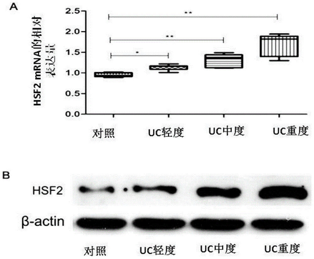
[0072] Example 3: Analysis of differential expression of HSF2 in mucosal tissues of UC patients with different degrees detected by real-time quantitative PCR and Western blot.
[0073] (1) Detection of differential expression of HSF2 by real-time fluorescent quantitative PCR
[0074] 1. Extraction of sample RNA
[0075] ① Take frozen and stored lysed intestinal mucosal cells, and let them dissolve completely at room temperature for 5 minutes.
[0076] ②Two-phase separation Add 0.2ml of chloroform to every 1ml of TRIZOL reagent lysed sample, and cap the tube tightly. Shake the tube vigorously by hand for 15 seconds, and incubate at 15 to 30°C for 2 to 3 minutes. Centrifuge at 12000 rpm for 15 minutes at 4°C. After centrifugation, the mixed liquid will be divided into the lower red phenol chloroform phase, the middle layer and the upper layer of the colorless aqueous phase. The RNA was all partitioned into the aqueous phase. The volume of the upper layer of the aqueous phas...
PUM
 Abstract
Description
Claims
Application Information
 - R&D
- Intellectual Property
- Life Sciences
- Materials
- Tech Scout
- Unparalleled Data Quality
- Higher Quality Content
- 60% Fewer Hallucinations
Browse by: Latest US Patents, China's latest patents, Technical Efficacy Thesaurus, Application Domain, Technology Topic, Popular Technical Reports.
© 2025 PatSnap. All rights reserved.Legal|Privacy policy|Modern Slavery Act Transparency Statement|Sitemap|About US| Contact US: help@patsnap.com


