Preparation method and application of anti-HCMV (human cytomegalovirus) Pp65 protein monoclonal antibody

A monoclonal antibody and antigen technology, applied in the field of genetic engineering, can solve problems such as high price of diagnostic reagents

Inactive Publication Date: 2014-10-08
HUNAN NORMAL UNIVERSITY
View PDF2 Cites 7 Cited by
  • Summary
  • Abstract
  • Description
  • Claims
  • Application Information

AI Technical Summary

Problems solved by technology

At present, the key raw materials for the diagnosis of HCMV infection in my country mainly ...

Method used


Image

  • Preparation method and application of anti-HCMV (human cytomegalovirus) Pp65 protein monoclonal antibody
  • Preparation method and application of anti-HCMV (human cytomegalovirus) Pp65 protein monoclonal antibody
  • Preparation method and application of anti-HCMV (human cytomegalovirus) Pp65 protein monoclonal antibody

Examples


Embodiment 1

[0036] Example 1: Animal immunization and serum titer detection: Pp65 protein plantar plate immunization mice

[0037] The purified Pp65 protein was fully emulsified with complete Freund's adjuvant, and three BALB / c female mice aged 6-8 weeks were immunized with a dose of 100 μg each by plantar plate immunization method. Two weeks later, Pp65 recombinant protein was fully emulsified with Freund's incomplete adjuvant, and BALB / c mice were boosted with the same dose and method. The mouse serum was collected 1 day before each immunization, and the antibody titer in the serum was detected by indirect ELISA. Three days before the fusion, the plantar plates were immunized with Pp65 recombinant protein without adjuvant at a dose of 100 μg per mouse. The collected blood was diluted 1:5 in normal saline, and then the blood suspension was placed at 37°C for 2 hours, then moved to 4°C overnight, centrifuged at 4000 g for 10 minutes after hemagglutination, and the supernatant was absor...

Embodiment 2

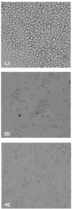
[0041] Example 2: Cell Fusion, Positive Hybridoma Screening and Subcloning

[0042] The SP2 / 0 myeloma cells in good growth state and lymph node cells of immunized mice were selected, washed three times with serum-free medium, mixed at 1:5~1:10, centrifuged at 1500 g for 5 min, and the supernatant was discarded. After gently shaking the centrifuge tube to disperse the cells evenly, slowly add 1 mL of 37 ℃ preheated PEG 1 500 and mix gently, then add 20 mL of 1640 medium. After centrifugation at 800 g for 5 min, discard the supernatant, add 20% FBS-HAT-1640 medium to suspend the cells and mix well.

[0043] The mixed cells were spread evenly in 96-well plates and placed at 37 °C, 5% CO 2 Cultured in an incubator, follow up and observe every day, change the medium with 10% FBS-HT-1640 culture medium 7-10 days after fusion, and detect with indirect ELISA when the clone grows to 1 / 3-1 / 2 of the bottom area of ​​the well For the antibody in the supernatant of hybridoma cells, th...

Embodiment 3

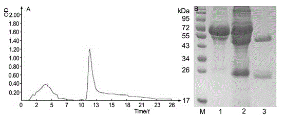
[0047] Example 3: Production and purification of monoclonal antibodies

[0048]The monoclonal antibody was produced by inducing ascites in mice. Take 8-10 w BALB / c mice, and inject sterile paraffin oil into the intraperitoneal cavity, 0.5 mL each. After 3 days, the 8D6 hybridoma cell line was injected into the abdominal cavity, and the cell density was adjusted to 1×10 6 ~2×10 6 / mL, each mouse was intraperitoneally injected with 0.5 mL of cell suspension. Seven to 12 days after cell inoculation, abdominal swelling of the mice could be clearly seen, and the ascites was collected and purified. The collected ascites was firstly purified by adding saturated ammonium sulfate precipitation at 1:1, centrifuged at 12,000 g for 10 min at 4°C, discarded the supernatant, and used a certain volume of PBS buffer (20 mmol / L NaCl, 2.68 mmol / L KCl, 10 mmol / L Na 2 HPO 4 , 1.76 mmol / L KH 2 PO 4 , pH 7.4) were dissolved, dialyzed twice into 10 times the volume of PBS buffer, 2 h each ...


PUM


Abstract

The invention discloses a high-specificity and high-affinity anti-Pp65 monoclonal antibody which is generated by a hybridoma cell strain 8D6 of the anti-Pp65 monoclonal antibody, wherein the cell strain 8D6 is collected in China Center For Type Culture Collection (CCTCC) and has a collection number of CCTCC NO:C201476. The monoclonal antibody secreted by the hybridoma cell strain has good affinity and specificity, and the immunoreactivity and the specificity of the antibody are superior to those of domestic and imported monoclonal antibodies. The anti-Pp65 monoclonal antibody can serve as a key raw material for blood white cell immunofluorescent cytochemical diagnosis and immunofluorescent quantitative PCR (polymerase chain reaction) detection of HCMV (human cytomegalovirus) infection, also can serve as a key raw material for detecting HCMV by using a double antibody sandwich method, has important meaning for establishment of a quick and sensitive clinical diagnosis reagent to HCMV infection, and has important application prospects.

Description

technical field [0001] The invention belongs to the field of genetic engineering, and in particular relates to a preparation method and application of an anti-HCMV Pp65 protein monoclonal antibody. Background technique [0002] Human cytomegalovirus (HCMV) is a double-stranded DNA virus with a nucleic acid size of about 235 kb, belonging to the genus β of the family Herpesviridae. HCMV infection is extremely common[1] and can cause severe disease in immunocompromised individuals and neonates. Most infected persons are asymptomatic, but maternal infection during pregnancy can lead to fetal infection, which can enter various target organs through the blood, resulting in multisystem Organ damage (such as pneumonia, hepatitis and encephalitis in a few cases), survivors are accompanied by neurological deafness, mental retardation and other neurological sequelae, so early diagnosis and treatment of HCMV infection is particularly important. HCMV Pp65 protein is a structural protei...

Claims


Application Information

Patent Timeline
IPC IPC(8): C07K16/08C12N5/20G01N33/577G01N33/569
Inventor 刘如石邱义兰许凤廖旻晶邱果李桑李烨刘胜姿
Owner HUNAN NORMAL UNIVERSITY
Why Eureka
Try Eureka