Nucleic acid detection method for separating luminous marker based on magnetic beads and nucleic acid hydrolysis
A technology of luminescent markers and detection methods, which is applied in the field of nucleic acid detection based on magnetic beads and nucleic acid hydrolysis to separate luminescent markers, and can solve problems such as limited detection sensitivity and weakened luminescent signal intensity
- Summary
- Abstract
- Description
- Claims
- Application Information
AI Technical Summary
Problems solved by technology
Method used
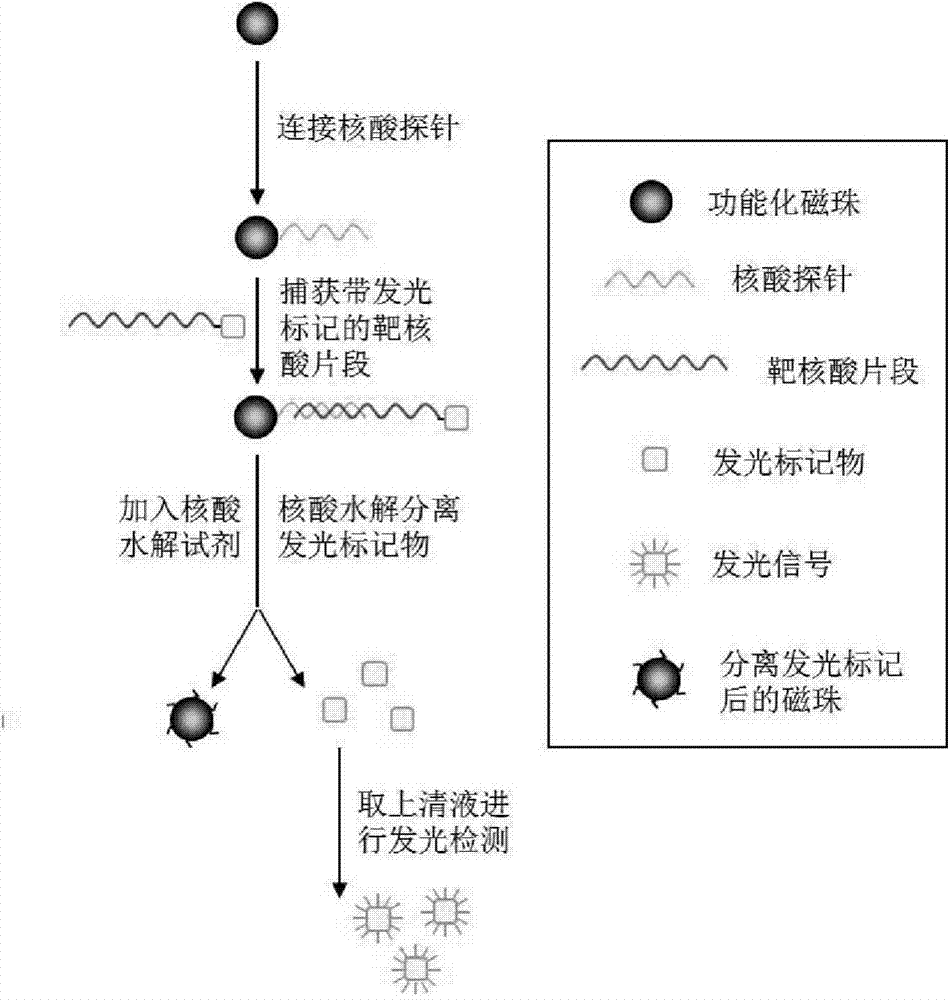
Image

Examples
Embodiment 1
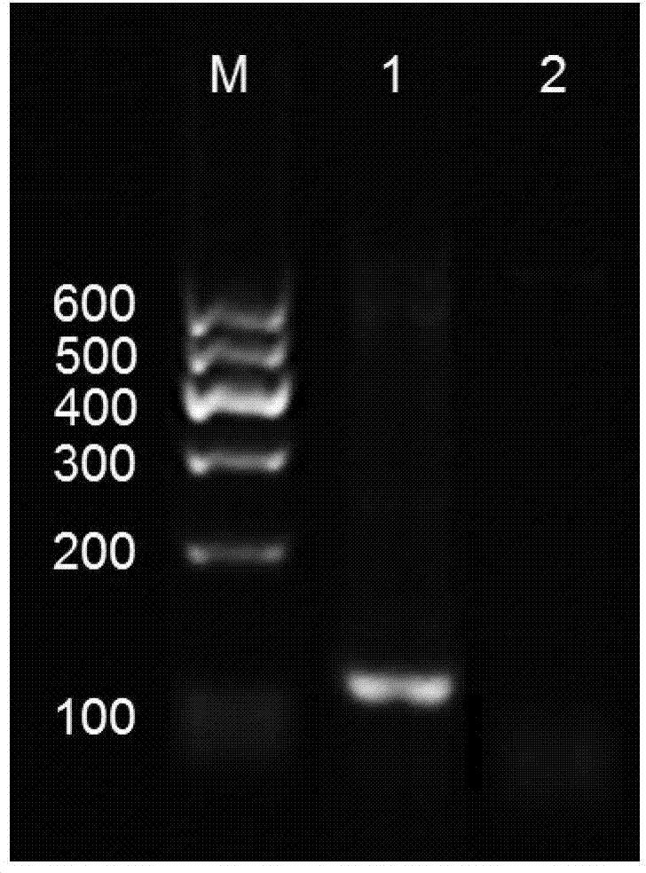
[0040] Embodiment 1 detects with hepatitis B virus (Hepatitis B virus, HBV) DNA as the object
[0041] 1. Carry out functional modification on the surface of magnetic beads: (1) Amination modification of magnetic beads: 1mL Fe 3 o 4 SiO 2 Magnetic beads (30 mg / mL) were magnetically separated, 6 mL of ethanol / water mixture was added and dispersed ultrasonically, then 12 μL of aminopropyltriethoxysilane (APTES) was added to the above mixture, and stirred at room temperature for 7 h. Use an external magnetic field to separate the APTES-modified magnetic particles, wash them with ethanol and dimethylformamide (N,N-dimethylformamide, DMF) several times, and disperse them in DMF at 5 mg / mL for later use; (2) magnetic Bead carboxylation modification: 1 mL of aminated magnetic beads (5 mg / mL) was added dropwise to an equal volume of succinic anhydride solution (succinic anhydride, SA, 1 mM) dissolved in DMF, and shaken at 37 ° C for 12 h. Wash several times with deionized water and...
Embodiment 2
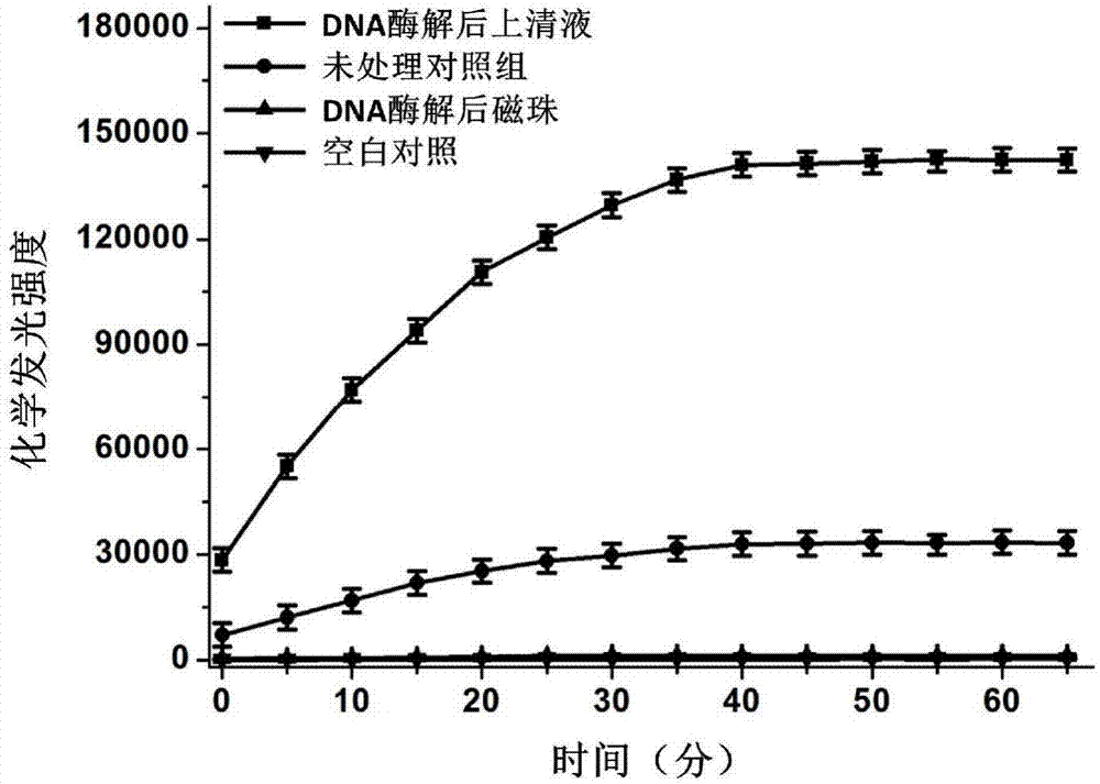
[0048] Example 2: Detection of serum miR-21 as an object
[0049] 1. Carry out functional modification on the surface of magnetic beads: the same method as described in Example 1 for functional modification on the surface of magnetic beads.
[0050] 2. Design a functional capture probe and a reporter probe that are complementary to the miR-21 sequence. The 3' end of the capture probe is modified with amino groups, and the 5' end of the reporter probe is modified with biotin. The probe sequences are as follows: 5'-CTGATAAGCTA-(T)15-NH2-3', 5'-Biotin-(T)15-TCAACATCAG-3'.
[0051] 3. Modification of capture probes on the surface of magnetic beads: Dilute carboxylated magnetic beads (5mg / mL) with an equal volume of 2-N-morpholinoethanesulfonic acid hydrate solution (2-(N-morpholino)ethanesulfonic acid hydrate, MES , 25mM, pH 6) Wash several times, discard the supernatant by magnetic separation, add the probe (1μM) diluted in MES solution to the washed carboxylated magnetic beads,...
PUM
 Abstract
Description
Claims
Application Information
 - R&D
- Intellectual Property
- Life Sciences
- Materials
- Tech Scout
- Unparalleled Data Quality
- Higher Quality Content
- 60% Fewer Hallucinations
Browse by: Latest US Patents, China's latest patents, Technical Efficacy Thesaurus, Application Domain, Technology Topic, Popular Technical Reports.
© 2025 PatSnap. All rights reserved.Legal|Privacy policy|Modern Slavery Act Transparency Statement|Sitemap|About US| Contact US: help@patsnap.com