Electric stimulation antibiosis dressing device

An electrical stimulation, conductive clip technology, applied in artificial respiration, physical therapy and other directions, to prevent the formation of bacterial biofilms, easy to operate, and promote wound healing.

Inactive Publication Date: 2015-04-22
SOUTHEAST UNIV
View PDF3 Cites 2 Cited by
  • Summary
  • Abstract
  • Description
  • Claims
  • Application Information

AI Technical Summary

Problems solved by technology

Although the drug resistance of biofilm bacteria is equivalent to 500-1000 times that of planktonic bacteria, the concentration required to kill biofilm bacteria is only 1.5-4 times that of the same type of planktonic bacteria by using the bioelectric effect

Method used


Image

  • Electric stimulation antibiosis dressing device

Examples


Embodiment 1

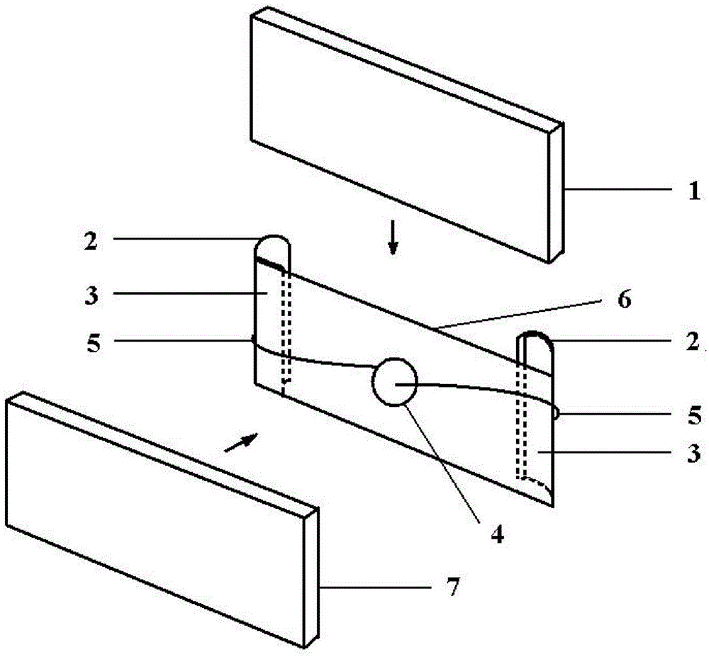
[0038] The bacterial cellulose membrane was obtained by fermentation of Acetobacter xylinum. After alkaline purification and freeze-drying, the membrane was fully immersed in a pyrrole monomer solution with a concentration of 5g / L, which contained 10μg / mL gentamicin. After 24 hours, place the film on a glass slide, apply direct current to both ends of the film, adjust the current to 0.25mA, and polymerize for 30 minutes. After the reaction was completed, the residual monomer solution on the surface was washed with deionized water. The conductivity of the conductive bacterial cellulose membrane is 0.05 S / cm. The prepared conductive bacterial cellulose dressing film 1 is fixed by insulating clips 2 and conductive clips 3, one side of the dressing film 1 is in contact with the liner 6, and the other side is exposed to the air. The conductive clips 3 located at both ends of the backing plate 6 are respectively in contact with and fixed on the front and back sides of the dressing ...

Embodiment 2

[0040]The bacterial cellulose membrane was obtained by fermentation with Agrobacterium, after alkaline purification and freeze-drying, the membrane was fully immersed in a pyrrole monomer solution with a concentration of 10g / L, which contained 10μg / mL amikacin. After 48 hours, place the film on a glass slide, apply direct current to both ends of the film, adjust the current to 0.35mA, and polymerize for 15 minutes. After the reaction was completed, the residual monomer solution on the surface was washed with deionized water. The prepared conductive bacterial cellulose dressing film 1 is fixed by insulating clips 2 and conductive clips 3, one side of the dressing film 1 is in contact with the liner 6, and the other side is exposed to the air. The conductive clips 3 located at both ends of the backing plate 6 are respectively in contact with and fixed on the front and back sides of the dressing film 1 . The conductive clip 3 is connected to the positive and negative poles of th...

Embodiment 3

[0042] The bacterial cellulose membrane was fermented by Acetobacter xylinum. After alkaline purification and freeze-drying, the membrane was fully immersed in a pyrrole monomer solution with a concentration of 5 g / L, which contained 5 μg / mL kitasamycin. After 10 hours, place the film on a glass slide, apply direct current to both ends of the film, adjust the current to 0.20 mA, and polymerize for 30 minutes. After the reaction was completed, the residual monomer solution on the surface was washed with deionized water. The prepared conductive bacterial cellulose dressing film 1 is fixed by insulating clips 2 and conductive clips 3, one side of the dressing film 1 is in contact with the liner 6, and the other side is exposed to the air. The conductive clips 3 located at both ends of the backing plate 6 are respectively in contact with and fixed on the front and back sides of the dressing film 1 . The conductive clip 3 is connected to the positive and negative poles of the butt...


PUM


Abstract

The invention relates to an electric stimulation antibiosis dressing device which comprises a dressing film (1), a pair of insulation clamping pieces (2) arranged oppositely, conducting clamping pieces (3) arranged on the insulation clamping pieces (2) respectively, a button cell (4), lead wires (5), a lining plate (6) which is arranged between the insulation clamping pieces (2) and are connected with the insulation clamping pieces (2) and an outer shell (7). The dressing film (1) is fixed by the insulation clamping pieces (2) and the conducting clamping pieces (3), one face of the dressing film (1) is in contact with the lining plate (6), and the other face of the dressing film (1) is in contact with a skin wound surface. The conducting clamping pieces (3) placed at the two ends of the lining plate (6) are in contact with the front face and the back face of the dressing film (1) respectively in a fixing mode. The conducting clamping pieces (3) are connected with a positive electrode and a negative electrode of the button cell (4) through the lead wires (5). The button cell (4) is fixed on the lining plate (6) and is covered by the outer shell (7). The electric stimulation antibiosis dressing device is simple in design and convenient to operate, the wound surface is subjected to contact treatment, bacterial biofilm forming can be effectively avoided, and wound healing is promoted.

Description

technical field [0001] The invention relates to an electric stimulation antibacterial dressing device, which belongs to the field of biomedical materials and related fields. Background technique [0002] After trauma, the skin, as the natural barrier against microbial invasion, is destroyed and prone to bacterial infection. Planktonic bacteria form biofilm in a short period of time, and bacterial biofilm releases planktonic bacteria intermittently, leading to repeated, aggravated or acute attacks of infection. [0003] Conventional drugs can kill planktonic bacteria and surface bacteria, and the clinical symptoms can be controlled, but they generally cannot penetrate bacterial biofilms. Studies have shown that the ability of the same bacteria to resist antibiotics and fungicides in a biofilm is 500-5000 times higher than that in a planktonic state. Due to this resistance to antibacterial drugs, bacterial biofilms exist in the host for a long time. When the environment permi...

Claims


Application Information

Patent Timeline
Patent Type & Authority Applications(China)
IPC IPC(8): A61N1/36
Inventor 姚琛周宾
Owner SOUTHEAST UNIV
Why Eureka
Try Eureka