Medical cosmetic product containing autologous peripheral blood vascular endothelial progenitor cells
A technology for endothelial progenitor cells and cosmetic products, which is applied in the field of medical cosmetic products containing autologous peripheral blood endothelial progenitor cells, can solve problems such as unacceptable, trauma, pain, etc., and achieves promotion of proliferation and survival, improvement of blood supply, and promotion of blood vessels. effect of growth
- Summary
- Abstract
- Description
- Claims
- Application Information
AI Technical Summary
Problems solved by technology
Method used
Image
Examples
preparation example 1
[0041] Preparation Example 1 Preparation of platelet-rich plasma
[0042] Use two 50ml syringes to extract 2ml of 1000U / ml heparin (Hebei Changshan Biochemical Pharmaceutical Co., Ltd.), collect 100ml of anticoagulant meridian blood, shake well, and add the collected heparin anticoagulant blood into two 50ml sterile centrifuge tubes In the process, centrifuge at a speed of 1000 rpm for 10 minutes, use a 5ml pipette to absorb the upper layer of plasma after centrifugation, and place it in a new 50ml centrifuge tube to obtain about 40ml of autologous plasma. The autologous plasma was centrifuged again at 3000 rpm for 10 minutes to obtain about 40ml of platelet-rich plasma, which was divided into 15ml sterile centrifuge tubes and stored in a 4°C refrigerator for later use.
preparation example 2
[0043] Preparation Example 2 Preparation of Autologous Endothelial Progenitor Cells
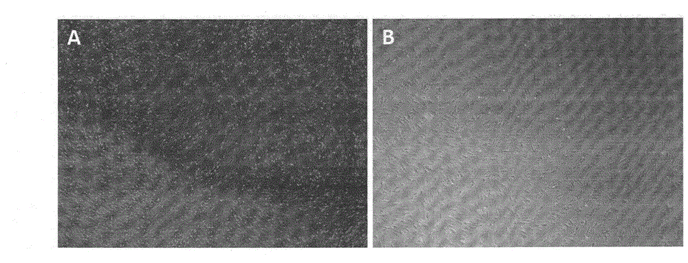
[0044] Take 60ml of blood cells obtained from autologous plasma by centrifugation in Preparation Example 1, dilute with 60ml of 1×PBS (pH 7.4), add to Ficoll density separation medium, and undergo density gradient centrifugation at 1800rpm for 30min to obtain mononuclear cells. Count under an inverted microscope. Endothelial cell culture medium (EGM-2) (containing 2% fetal bovine serum, 10 U / ml heparin, 12 μg / ml bovine brain extract, 10 ng / ml recombinant human epidermal growth factor, 1 μg / ml hydrocortisone) for monocytes after counting Pine, 50ng / ml recombinant human vascular endothelial growth factor, 50ng / ml recombinant human R3-insulin-like growth factor-1 and 50ng / ml recombinant human basic fibroblast growth factor-β) resuspended, press 5×10 6 Cells / ml were added to recombinant human fibronectin-coated 25cm 2 5ml in culture flask, at 37°C, 5% CO 2 Grow in the incubator for 48 hours. ...
Embodiment 3
[0048] Example 3 Flow Cytometry Identification of Autologous Endothelial Progenitor Cells
[0049] Take the 2 × 10 samples prepared in Preparation Example 1 after trypan blue staining and counting. 6Endothelial progenitor cells were divided into two groups. The first group was added with 20 μL FITC-labeled mouse anti-human FLK-1 monoclonal antibody, 20 μL PE-labeled mouse anti-human CD133 monoclonal antibody and 20 μL Percp-labeled mouse anti-human CD34 monoclonal antibody; the second group As an isotype control, add 20 μL FITC-labeled mouse IgG1, 20 μL PE-labeled mouse IgG1 and 20 μL PerCP-labeled mouse IgG1 respectively; place in 4°C refrigerator for staining for 30 minutes, then wash three times with 1 mL of 1× phosphate buffer, and finally wash with The washed cells were resuspended in 0.5 mL of 1×PBS, and the washed cells were detected by a FACSCalibur clinical flow cytometer. figure 2 As detected by flow cytometry, the endothelial progenitor cells prepared in Preparati...
PUM
 Abstract
Description
Claims
Application Information
 - R&D
- Intellectual Property
- Life Sciences
- Materials
- Tech Scout
- Unparalleled Data Quality
- Higher Quality Content
- 60% Fewer Hallucinations
Browse by: Latest US Patents, China's latest patents, Technical Efficacy Thesaurus, Application Domain, Technology Topic, Popular Technical Reports.
© 2025 PatSnap. All rights reserved.Legal|Privacy policy|Modern Slavery Act Transparency Statement|Sitemap|About US| Contact US: help@patsnap.com


