Method for preparing decellularized lamellar cornea matrix sheet

A technology of lamellar cornea and corneal stroma, applied in the field of biomedical tissue engineering, can solve the problem of biocompatibility, strength, degradation rate, transparency, homology, antigenic and pathogenic defects, and easily damaged tissue structure , affect the transparency of the cornea and other issues, to achieve the effect of simple and reliable production process, complete decellularization, and low cost

Inactive Publication Date: 2015-05-27
南昌大学第一附属医院 +1
View PDF1 Cites 12 Cited by
  • Summary
  • Abstract
  • Description
  • Claims
  • Application Information

AI Technical Summary

Problems solved by technology

However, various seed cell scaffold materials developed at home and abroad have defects in corneal curvature and topography, biocompatibility, strength, degradation rate, transparency, homology, antigenicity and pathogenicity.
It is considered that the most promising research is the heterogeneous corneal stroma that has been decellularized. However, the commonly used decellularization method is cumbersome, complicated, and easy to damage the tissue structure, which seriously affects the transparency of the cornea and makes the effect poor.

Method used


Image

  • Method for preparing decellularized lamellar cornea matrix sheet
  • Method for preparing decellularized lamellar cornea matrix sheet
  • Method for preparing decellularized lamellar cornea matrix sheet

Examples


preparation example Construction

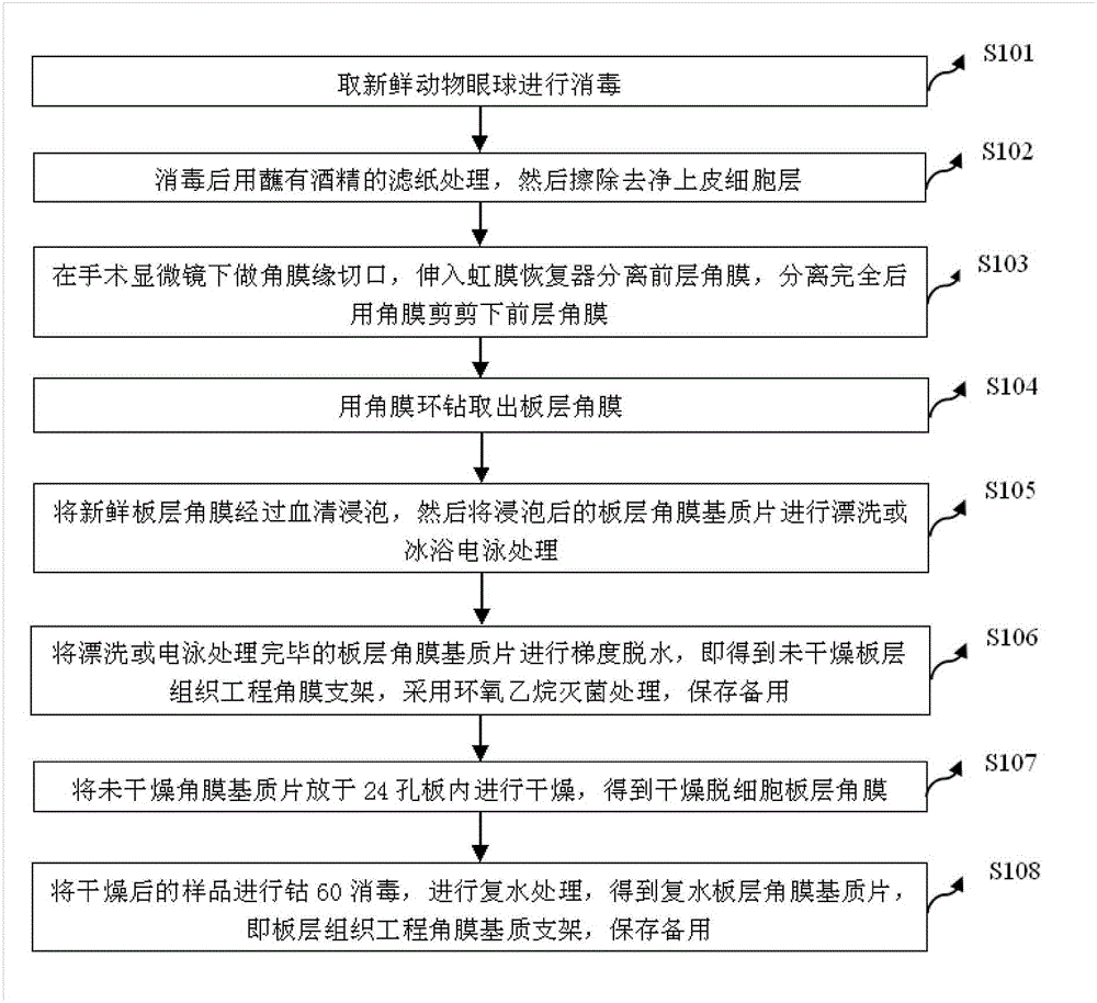
[0035] figure 1 The implementation flow of the method for preparing the decellularized lamellar corneal stromal sheet provided by the embodiment of the present invention is shown. The method includes the following steps:

[0036] In step S101, fresh animal eyeballs are taken for disinfection;

[0037] In step S102, after disinfection, it is treated with filter paper dipped in alcohol, and then the epithelial cell layer is wiped off;

[0038] In step S103, make a limbal incision under the operating microscope, insert the iris restorer to separate the anterior cornea, and cut off the anterior cornea with corneal scissors after the separation is complete;

[0039] In step S104, the lamellar cornea is taken out with a corneal trephine;

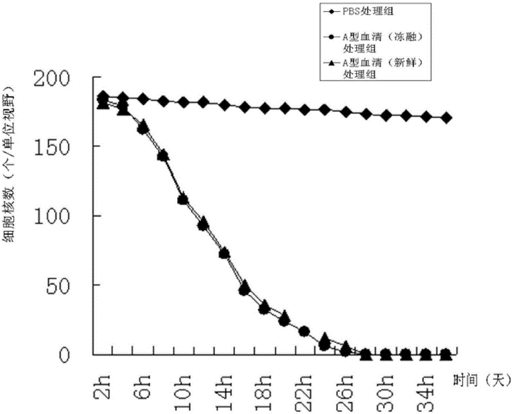
[0040] In step S105, the fresh lamellar cornea is soaked in serum, and then the soaked lamellar corneal stroma is rinsed or treated by ice bath electrophoresis;

[0041] In step S106, the lamellar corneal stroma sheet that has been rinsed or e...

Embodiment 1

[0052] Disinfect fresh animal eyeballs by immersing them in 0.5-2% iodophor for 2-5 minutes. After soaking, rinse them with PBS for 5 minutes each time; Cover the surface of the cornea with filter paper or soak it directly for 1-5min, then gently wipe the epithelial cell layer with a cotton swab / corneal epidermis knife; make a limbal incision with a jewel knife under the operating microscope, and insert the iris restorer to separate the anterior cornea After the separation is complete, use corneal scissors to cut off the anterior cornea. The limbal incision is about 1 / 3 deep, 0.2-0.8mm thick, and 3mm long. The thickness of the separated lamellar layer is 0.2-0.8mm, or directly use lamellar cornea Separate the anterior lamellar cornea about 1 / 3 deep (about 0.2-0.8mm thick) with a splitting knife; then put the separated anterior lamellar cornea on the corneal pillow (the anterior elastic layer faces down), and use a corneal trephine to remove 3-10mm diameter lamellar cornea; the...

Embodiment 2

[0054] Disinfect fresh animal eyeballs by immersing them in PBS containing antibiotics for 5-10 minutes. After soaking, wash three times with PBS for 5 minutes each time; after disinfection, cover the surface of the cornea with filter paper dipped in 15-30% alcohol or directly soak for 1-5 minutes, and then use a cotton swab / The corneal epidermis gently wipes off the epithelial cell layer; under the operating microscope, use a gemstone knife to make a limbal incision, insert the iris restorer to separate the anterior cornea, and cut off the anterior cornea with corneal scissors after the separation is complete. The limbal incision is deep About 1 / 3, the thickness is 0.2-0.8mm, the length is 3mm, the thickness of the separated lamellar layer is 0.2-0.8mm, or the depth of about 1 / 3 (thickness is about 0.2-0.8mm) is directly separated with a lamellar keratectomy knife Anterior lamellar cornea; then put the separated anterior lamellar cornea on the corneal pillow (the anterior el...


PUM

PropertyMeasurementUnit
thicknessaaaaaaaaaa

Abstract

The invention discloses a method for preparing a decellularized lamellar cornea matrix sheet. The method comprises the steps of disinfecting a fresh animal eyeball; treating with filter paper dipped with alcohol after disinfecting, and then erasing an epithelial cell layer; incising the corneal limbus under an operation microscope, stretching into an iris restorer to separate the anterior cornea, and then shearing the anterior cornea with corneal scissors; drilling the lamellar cornea with a corneal annulus; soaking the fresh lamellar cornea into serum, and then rinsing or performing ice-bath electrophoresis treatment; performing gradient dehydration after the treatment to obtain a non-dried lamellar tissue engineering corneal frame, sterilizing with ethylene oxide, and preserving for later use; drying the non-dried cornea matrix sheet in a 24-pore plate to obtain dried decellularized lamellar cornea; and sterilizing the dried sample with cobalt 60, and performing rehydration treatment to obtain a rehydrated lamellar cornea matrix sheet. The obtained cornea matrix sheet is high in transparency, low in structural destroy, good in biocompatibility, close to fresh cornea in performance and thorough in decellularization.

Description

technical field [0001] The invention belongs to the technical field of biomedical tissue engineering, and in particular relates to a preparation method of an acellular lamellar corneal stroma sheet. Background technique [0002] Corneal disease is one of the common blinding eye diseases. Clinically, corneal inflammation and corneal damage caused by various factors can cause corneal defect, erosion and ulcer. If not treated effectively, the visual function of the patient will be seriously damaged. and quality of life. According to the report of the World Health Organization, corneal disease is currently the second major cause of vision loss, second only to cataracts, and the number of new corneal blindness caused by corneal ulcers and eye trauma is 1.5 million to 2 million every year, which seriously threatens patients. health. The only effective treatment for corneal blindness is keratoplasty. However, due to the influence of religious beliefs and customs, very few people...

Claims


Application Information

Patent Timeline
Patent Type & Authority Applications(China)
IPC IPC(8): A61L27/36A61L27/50
Inventor 邵毅
Owner 南昌大学第一附属医院
Why Eureka
Try Eureka