
Method for testing ion transmission condition of peptide fragment in simulated environment

A technology of ion transmission and simulated environment, used in material excitation analysis, fluorescence/phosphorescence, etc., can solve the problems of high requirements for instruments and operators, high professionalism, complex operation, etc., and achieves low professional requirements, short time-consuming, Simple to use effects

Active Publication Date: 2015-06-24
宁波市博坤生物科技有限公司
View PDF4 Cites 0 Cited by
  • Summary
  • Abstract
  • Description
  • Claims
  • Application Information

AI Technical Summary

Problems solved by technology
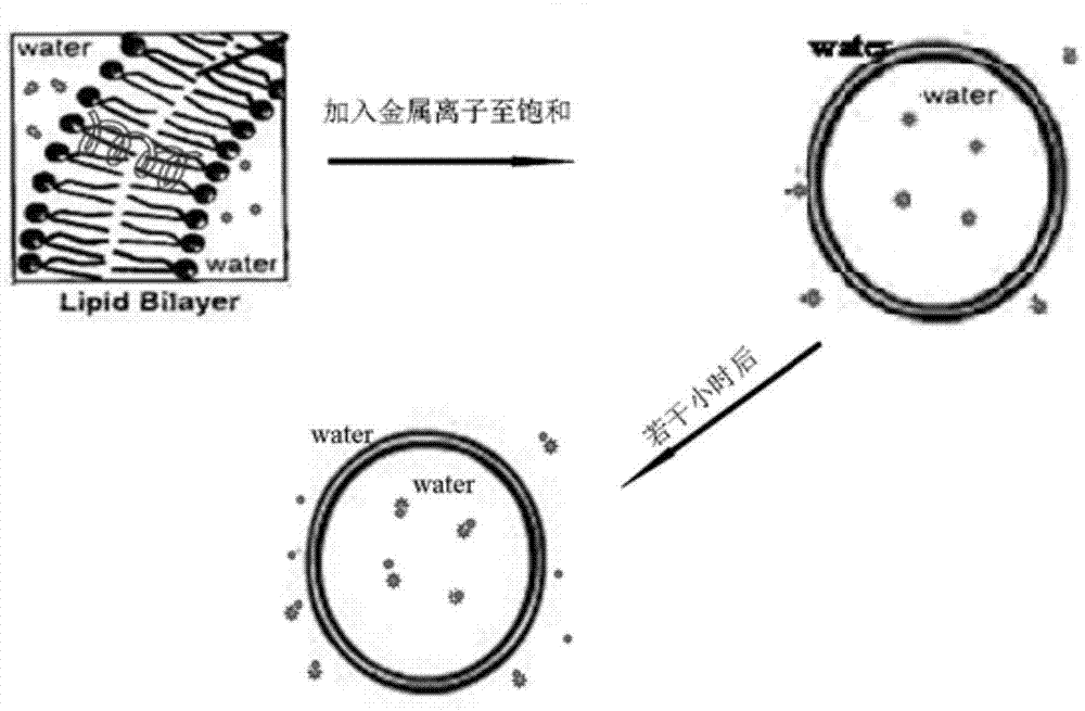
[0007] In order to solve the above technical problems, the present invention uses fluorescence spectroscopy to test the ion transmission of peptides in a simulated environment, which solves the complex operation, high professionalism, high cost, and relatively high requirements for instruments and operators in the prior art. high disadvantage

Method used


Image

  • Method for testing ion transmission condition of peptide fragment in simulated environment
  • Method for testing ion transmission condition of peptide fragment in simulated environment
  • Method for testing ion transmission condition of peptide fragment in simulated environment

Examples


Embodiment 1

[0037] Step 1: Prepare a buffer solution: prepare 200 ml of a disodium hydrogen phosphate-citric acid buffer solution with a pH value of 3 with deionized water, and add 0.500 g of sodium chloride;

[0038] Step 2: Prepare the standard sample stock solution of chlortetracycline: Accurately weigh 0.501 mg of standard sample of chlortetracycline with an electronic balance, use ultrapure water to make up to 10 mL in a brown volumetric flask, dissolve it by ultrasonic, and prepare 0.05 g / L stock solution, sealed and placed in a 4°C refrigerator for later use;

[0039] Step 3: Preparation of phospholipid unilamellar vesicles without added peptides: put 0.5 mg of dimyristoylphosphatidylglycerol into a sample tube, add 80 μl of a mixed solvent of chloroform and methanol at a volume ratio of 2:1, and wait for dimyristoyl phosphatidylglycerol to After the acylphosphatidylglycerol is completely dissolved, nitrogen gas is blown in until the solvent evaporates completely, and the liposome ...

Embodiment 2

[0047] Step 1: prepare a buffer solution: prepare 250 ml of a disodium hydrogen phosphate-citric acid buffer solution with a pH value of 7 with deionized water, and add 0.731 g of sodium chloride;

[0048] Step 2: Prepare the standard stock solution of chlortetracycline: Accurately weigh 2.000 mg of the standard sample of chlortetracycline with an electronic balance, use ultrapure water to make up to 10 mL in a brown volumetric flask, dissolve it with ultrasonic waves, and prepare 0.2 g / L stock solution, sealed and placed in a 4°C refrigerator for later use;

[0049] Step 3: Preparation of phospholipid unilamellar vesicles without added peptides: put 0.8 mg of dimyristoylphosphatidylglycerol into a sample tube, add 90 μl of a mixed solvent of chloroform and methanol at a volume ratio of 2:1, and wait until the After the myristoylphosphatidylglycerol is completely dissolved, blow in nitrogen until the solvent evaporates completely. The liposome forms a transparent film at the b...

Embodiment 3

[0059] Step 1: Prepare a buffer solution: prepare 250 ml of a disodium hydrogen phosphate-citric acid buffer solution with a pH value of 4 with deionized water, and add 0.731 g of sodium chloride;

[0060] Step 2: Prepare the standard stock solution of chlortetracycline: Accurately weigh 1.000 mg of the standard sample of chlortetracycline with an electronic balance, use ultrapure water to make up to 10 mL in a brown volumetric flask, dissolve it by ultrasonic, and prepare 0.1 g / L stock solution, sealed and placed in a 4°C refrigerator for later use;

[0061] Step 3: Preparation of phospholipid unilamellar vesicles without added peptides: put 1.2 mg of dimyristoylphosphatidylglycerol into a sample tube, add 100 μl of a mixed solvent of chloroform and methanol at a volume ratio of 2:1, and wait for dimyristoyl phosphatidylglycerol to After the acylphosphatidylglycerol is completely dissolved, nitrogen gas is blown in until the solvent is completely evaporated, and the liposome ...


PUM

PropertyMeasurementUnit
Wavelengthaaaaaaaaaa
Excitation wavelengthaaaaaaaaaa

Abstract

The invention belongs to the technical field of biology and particularly relates to a method for testing an ion transmission condition of a peptide fragment in a simulated environment. The testing method is a fluorescent spectrometry; and ions are metal ions. The testing method comprises the following six steps: preparing a buffering solution; preparing a standard sample stock solution of aureomycin, preparing a phospholipid monolayer vesicle without the peptide fragment, carrying out fluorescence detection on the phospholipid monolayer vesicle without the peptide fragment, preparing a phospholipid monolayer vesicle with the peptide fragment, and carrying out the fluorescence detection on the phospholipid monolayer vesicle with the peptide fragment. Aiming at the peptide fragment of a fourth membrane-spanning protein region, a testing method and a testing condition are further improved; by virtue of the testing method, the defects that the operation is complicated, the specialty is high, the cost is high and the requirements on instruments and operators are relatively high in the prior art are overcome; and the ion transmission condition of the fourth membrane-spanning protein region can be relatively conveniently researched, and a transmission function of a whole solute transportprotein transmission body is achieved.

Description

technical field [0001] The invention belongs to the field of biotechnology, and in particular relates to a method for testing the ion transmission of peptide segments in a simulated environment. Background technique [0002] Genome sequencing data revealed that a quarter of the proteins deciphered in DNA were membrane proteins. Membrane proteins are a class of proteins with a special structure, which are embedded in phospholipid membranes and are at the interface between cells and the outside world. Membrane proteins are basically divided into two categories: peripheral proteins and internal proteins. Membrane proteins that run through the entire phospholipid bilayer membrane are also called transmembrane proteins. Membrane proteins control the basic activities of many living organisms, such as cell growth, division and apoptosis, the transmission of various signals between cells, and the sense of smell and taste of living organisms. Membrane proteins form receptors for va...

Claims


Application Information

Patent Timeline
IPC IPC(8): G01N21/64
Inventor 齐海燕翟明翚王颖苏立强张晓红刘晓兰祖广权
Owner 宁波市博坤生物科技有限公司
Learn More