
A separation culture method for lens epithelium stem cells

A technology for separating and culturing lenses, applied in non-embryonic pluripotent stem cells, animal cells, vertebrate cells, etc., to achieve the effects of easy operation, good application prospects and high activity

Active Publication Date: 2015-07-22
GUANGZHOU KANGRUI BIOLOGICAL PHARMA TECH CO LTD
View PDF6 Cites 4 Cited by
  • Summary
  • Abstract
  • Description
  • Claims
  • Application Information

AI Technical Summary

Problems solved by technology

lens epithelial stem cells

Method used


Image

  • A separation culture method for lens epithelium stem cells
  • A separation culture method for lens epithelium stem cells
  • A separation culture method for lens epithelium stem cells

Examples


Embodiment 1

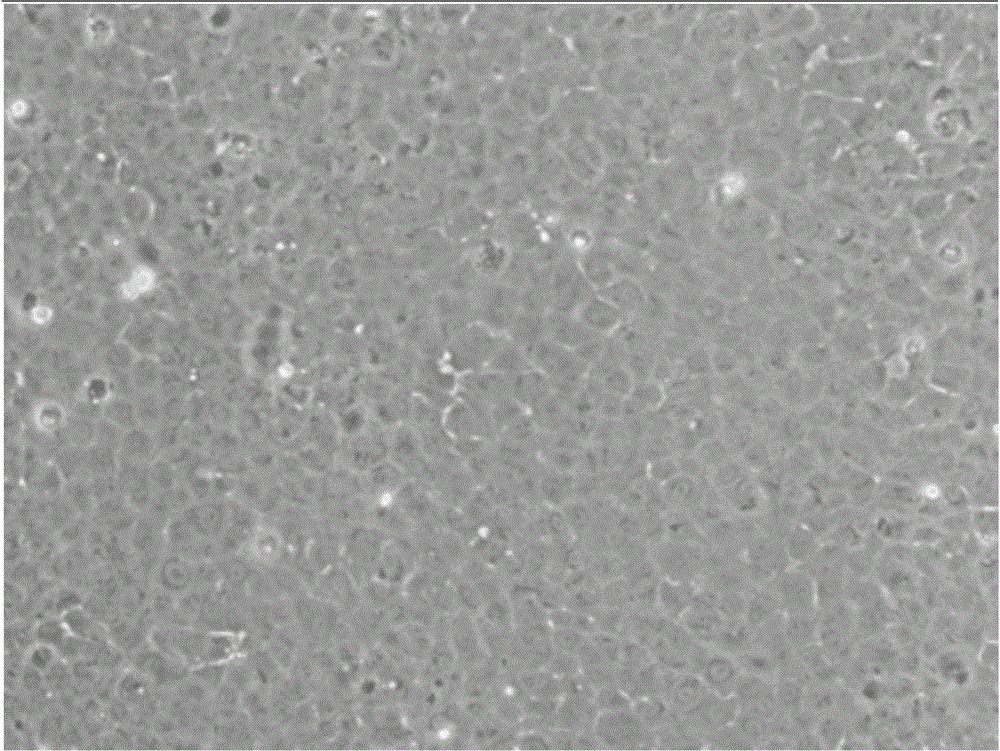
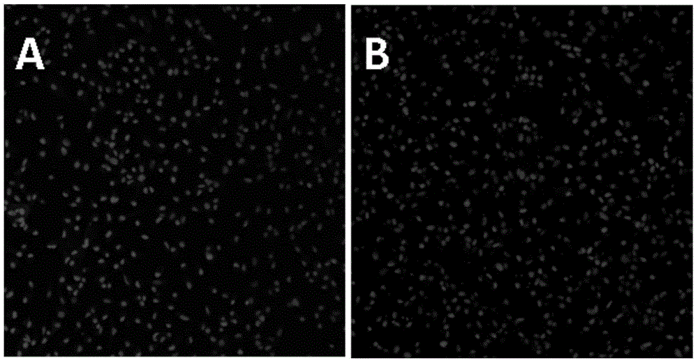
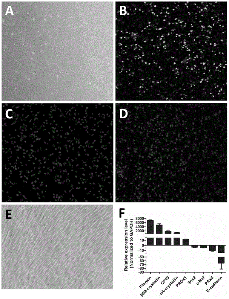
[0038] Example 1 Isolation, cultivation and identification of human eye lens epithelial stem cells

[0039] 1. Method

[0040] 1.1 Isolation and culture

[0041](1) The six-well plate was coated with DMEM / F12 culture solution containing 2% Matrigel (Matrigel is Matrigel) for 1 hour before use, the culture solution was discarded, and washed 5 times with sterile PBS;

[0042] (2) Rinse the eye tissue 3 times with PBS containing penicillin / streptomycin, cut off the cornea circularly in the ultra-clean table, cut and tear off the iris radially, fully expose the equator of the lens, and tear off the lens as close to the equator as possible with ophthalmic forceps Anterior capsule and attached epithelium, cut into 1×1mm 2 The fragments were added to a centrifuge tube containing 5 ml of 0.2% collagenase IV to obtain a cell mass. Then put the centrifuge tube into a 37°C incubator and shake gently for 2 hours for digestion. After 2 hours, centrifuge the cells at 1000 rpm for 5 minu...

Embodiment 2

[0056] Example 2 Isolation, Culture and Identification of Rabbit Eye Lens Epithelial Stem Cells

[0057] 1. Cell isolation and culture

[0058] All animal studies were performed in full compliance with the ARVO statement and relevant approvals for animals for ophthalmic and vision research. Eyeballs were removed from New Zealand white rabbits and washed 3 times with PBS containing antibiotics. After the cornea and iris are removed, a small incision is made behind the lens capsule, and the anterior lens capsule and attached epithelium are removed and cut into 1×1mm 2 , the fragments were placed in a centrifuge tube with 5ml of 0.2% collagenase IV to obtain a cell mass. Then put the centrifuge tube into a 37°C incubator and shake gently for 2 hours for digestion. After 2 hours, centrifuge the cells at 1000 rpm for 5 minutes at room temperature to vacuum-aspirate the collagenase. Add 5 ml of 0.25% trypsin-EDTA and pass through a 100um cell strainer. Filtered cells were cultu...


PUM


Abstract

A separation culture method for lens epithelium stem cells is disclosed. The method includes following steps: (1) cutting a lens anterior capsule into small pieces, digesting, and paving the obtained cells to a culture flask coated with a medium, a culture plate coated with a medium or a culture dish coated with a medium; and (2) adding an MEM culture solution containing 15-25% of FBS, 50-150 mug / L of a human epidermal growth factor (FGF), 5-10 mg / L of insulin, 5-10 mg / L of hydrocortisone, 5-10 mg / L of a vibrio cholera toxin, 0.01-0.05 mg / L of 3,3',5-iodo-L-para-hydroxyphenylalanine, 1-5 mg / L of adenine, 5-10 mg / L of glycine, 6-12 mg / L of alanine, 10-16 mg / L of asparagines, 10-16 mg / L of aspartic acid, 12-18 mg / L of glutamic acid, 8-15 mg / L of proline and 7-14 mg / L of serine, putting into an incubator having a temperature of 37 DEG C, culturing for 2-3 days to form clone, and forming lens stem cells with monolayer activity after 10-14 days. A separation culture solution for the lens epithelium stem cells is also disclosed. The method can separate and obtain the primary lens epithelium stem cells. The stem cells are high in activity. The method is simple, convenient, easy in operation and good in application prospect.

Description

technical field [0001] The invention relates to a method for separating and culturing lens epithelial stem cells. Background technique [0002] Stem cells are a type of pluripotent cells with self-replicating ability. Under certain conditions, it can differentiate into a variety of functional cells. Stem cells are divided into embryonic stem cells and adult stem cells according to their developmental stage. According to the developmental potential of stem cells, they are divided into three categories: totipotent stem cells, pluripotent stem cells and unipotent stem cells. Stem cells are not fully differentiated and immature cells, which have the potential to regenerate various tissues and organs and the human body. They are called "universal cells" in the medical field. [0003] The lens is a double-convex lens-shaped transparent tissue suspended behind the iris and directly in front of the vitreous by suspensory ligaments. It is an important refractive medium in the eye....

Claims


Application Information

Patent Timeline
IPC IPC(8): C12N5/074
Inventor 侯睿蔡惠民
Owner GUANGZHOU KANGRUI BIOLOGICAL PHARMA TECH CO LTD
Learn More