
New application of dcf1 gene
A 1.dcf1, gene technology, applied in gene therapy, nervous system diseases, genetic material components, etc., can solve the problems of no treatment for demyelination diseases, unclear mechanism of demyelination and remyelination, etc.
- Summary
- Abstract
- Description
- Claims
- Application Information
AI Technical Summary
Problems solved by technology
Method used
Image

Examples
Embodiment 1
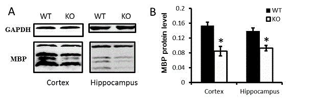
[0012] Example 1: The target area, corpus callosum and hippocampus were taken out of the pre-cooled PBS. Then homogenize in a homogenizer with Western and IP cell lysate until there is no obvious tissue block and it becomes a transparent homogenate. Continue to lyse on ice for 30 min, and then collect the lysate in a 1.5ml EP tube. The supernatant was collected by centrifugation at 8000 rpm for 10 minutes at 4°C. Continue to centrifuge the supernatant at 4°C, 15,000rpm for 20min, collect the supernatant again to be the tissue protein extract, add the corresponding amount of Loading buffer, heat and denature it for later use. Afterwards, Western blotting was performed. The electrophoresis conditions are stacking gel 80V, 20 minutes; separating gel 120V, 90 minutes. The transfer condition was 25V for 60 minutes. The primary antibody dilution ratio is 1:1000, and the secondary antibody dilution ratio is 1:10000. ( figure 1 A) Schematic of Western blotting (MBP / GAPDH) of the...
Embodiment 2
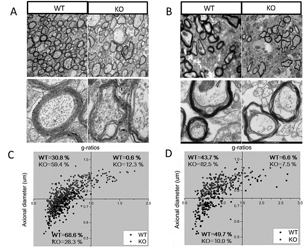
[0013] Example 2: Detection of myelin sheath morphology by transmission electron microscopy
[0014] Experimental mice were first perfused (4% paraformaldehyde and 1% glutaraldehyde in fixative). Then the mouse brain was taken out, placed in a fixative solution containing 2.5% glutaraldehyde, and the area to be observed (hippocampus, corpus callosum) was cut into 1mm 2 size and then fixed in the 2.5% glutaraldehyde fixative described above. This was followed by post-fixation with osmium tetroxide for 2 hours. Alcohol gradually dehydrates. Finally, the tissue block was embedded in epoxy resin. Slice with an ultra-thin microtome. Slices with a thickness of 500nm are placed on glass slides and stained with toluidine blue, and can be observed under Confocal; Next observe. ( figure 2 A, B) Schematic diagram of ultrastructural electron microscopy of myelin in the corpus callosum and hippocampus. It can be seen that in both the corpus callosum and hippocampus, the myelin she...
Embodiment 3
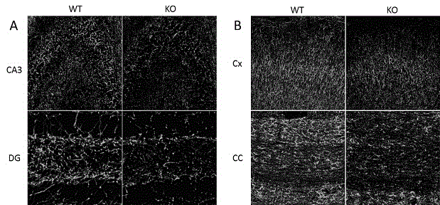
[0015] Example 3: Immunohistochemical detection
[0016] Firstly, the experimental mice were perfused and fixed with 4% PFA, dehydrated with sucrose, and embedded. Then cryosection (20um) was performed. Finally, immunohistochemistry was performed. Wash 3 times with 0.01M PBS, 5 minutes each; Permeabilize with 0.1%TritonX-100-PBS for 30 minutes; Wash 3 times with 0.01M PBS, 5 minutes each; Block with 2% BSA-PBS for 1 hour at room temperature; Blot dry 2% After BSA-PBS, directly drop the primary antibody diluted in PBS (MBP, 1:500). Incubate at 37°C for 2 hours, or overnight at 4°C; blot dry the primary antibody, wash with 0.01M PBS 3 times, 5 minutes each time. Under dark conditions, add secondary antibody diluted in PBS (1:500 dilution ratio). Incubate at 37°C for 2 hours; blot dry the secondary antibody, add DAPI (1:1000) diluted in PBS, and incubate at room temperature for 10 minutes; blot dry DAPI, wash 3 times with 0.01M PBS, 5 minutes each time; seal with fluorescent ...
PUM
 Abstract
Description
Claims
Application Information