Multiple detection method for six phospholipid in complex sample

A complex sample and multiple detection technology, applied in the field of biological analysis, can solve the problems of affecting the air tightness and service life of the machine, severe peak shape diffusion, and long separation time, and achieve good separation, high repeatability, and simple operation.

Active Publication Date: 2015-09-23
卢航 +1
View PDF2 Cites 10 Cited by
  • Summary
  • Abstract
  • Description
  • Claims
  • Application Information

AI Technical Summary

Problems solved by technology

The former has a better application in detection practice, but the polymer accessories in high performance liquid chromatography may swell or shrink in chloroform, which affects the airtightness and service life of the machine
The latter system i

Method used


Image

  • Multiple detection method for six phospholipid in complex sample
  • Multiple detection method for six phospholipid in complex sample
  • Multiple detection method for six phospholipid in complex sample

Examples


Embodiment 1

[0052] Embodiment 1. standard sample detection and standard curve preparation

[0053](1) Standard samples: PE (P7943), PI (P2517), PS (P7769), PC (P3556), SM (S0756) and LPC (L0906) standard products were purchased from Sigma-Aldrich.

[0054] (2) HPLC-ELSD analysis method:

[0055] Hitachi LaChrom Elite series high performance liquid chromatography;

[0056] Alltech2000 evaporative light detector, split mode, using air as atomizing gas, atomizing gas flow rate 1.7L / min; drift tube temperature 45°C;

[0057] Performance-Si type normal phase silica gel chromatography column, 100mm×4.6mm, column temperature 25℃;

[0058] The elution flow rate is 1.5mL / min, and the mobile phase of gradient elution is:

[0059] 0.0min, A 40%, B 57%, C 3%;

[0060] 8.0min, A 40%, B 50%, C 10%;

[0061] 15.0min, A 40%, B 50%, C 10%;

[0062] 15.1min, A 40%, B 57%, B 3%;

[0063] 24.0min, A 40%, B 57%, C 3%;

[0064] Wherein, mobile phase A is n-hexane containing 0.04% triethylamine, mobil...

Embodiment 2

[0070] The analysis method evaluation of embodiment 2.HPLC-ELSD

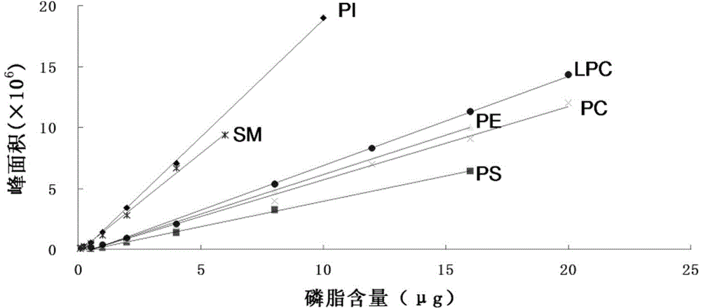
[0071] According to the detection method of Example 1, the mean value, RSD and C.V. value of the peak area of ​​three repeated injections with phospholipid standard. The results are shown in Table 2. Visible, the peak area reproducibility of all phospholipid standard samples is very good, and C.V. (coefficient of variation) value is all less than 3.5%.

[0072] Table 2. Reproducibility of peak areas of phospholipid standards

[0073] Phospholipid Standards

[0074] PC

Embodiment 3

[0075] Example 3. Based on HPLC-ELSD multiple detection of the composition and content of Apostichopus japonicus adenophospholipids

[0076] (1) Prefabricated samples to be tested:

[0077] The sea cucumber gonad sample tissue to be tested was thawed in running water, and homogenized using a high-speed tissue homogenizer. Mix it with chloroform-methanol (2:1, v / v) solution at a ratio of 1:10 (g / ml), stir, let stand for ten minutes, then filter with suction, collect the filtrate, dehydrate and spin dry to obtain total gonad lipid of sea cucumber. Weigh about 600 mg of sea cucumber gonad total fat, dissolve it in a small amount of chloroform, load the sample, and perform silica gel column chromatography. The neutral lipids were eluted successively with 5 times column volume of chloroform, the glycolipids and pigments were eluted with 3 times column volume of acetone, and the phospholipids were eluted with methanol. The phospholipids were dissolved in chloroform and stored froz...


PUM


Abstract

The invention discloses a multiple detection method for six phospholipid in a complex sample. The multiple detection method includes HPLC analysis, wherein specification of a positive-phase silica gel chromatography column is 100mmx4.6mm, column temperature of 25 DEG C, elution flow speed is 1.5mL/min, flowing phase of gradient elution includes 40% of A, 57% of B and 3% of C of 0.0min, 40% of A, 50% of B and 10% of C of 8.0min, 40% of A, 50% of B and 10% of C of 15.0min, 40% of A, 57% of B and 3% of C of 15.1min and 40% of A, 57% of B and 3% of C of 24.0min, flowing phase A is normal hexane containing triethylamine of 0.01-0.08%, flowing phase B is isopropanol, and flowing phase C is acetic acid solution of 10-20%. The multiple detection method is simple in operation, quick, accurate, good in component separating effect, high in repeatability and easy for application and popularization in detection practice.

Description

technical field [0001] The invention belongs to the field of biological analysis, in particular to the simultaneous detection of multiple phospholipid components in complex biological samples. Background technique [0002] There are many kinds of phospholipids in animals and plants, mostly phosphatidylcholine and phosphatidylethanolamine. Limited by the interference of complex components in biological samples, the detection of phospholipid components, especially the multiple detection methods of phospholipid components, needs to be developed urgently. [0003] In the prior art, commonly used methods for phospholipid analysis include thin-layer chromatography (TLC), high-performance liquid chromatography (HPLC), mass spectrometry (MS) and gas chromatography-mass spectrometry (C-MS) techniques, etc. With the development of mass spectrometry, more powerful LC-MS methods are also used in the targeted quantitative analysis of phospholipids. In detection practice, high performan...

Claims


Application Information

Patent Timeline
IPC IPC(8): G01N30/02G01N30/06
Inventor 卢航胡建恩武龙赵慧陈慧民李晶晶
Owner 卢航
Why Eureka
Try Eureka