
Preparation method of tissue engineering cornea

A technology of tissue engineering and cornea, which is applied in the field of tissue engineering cornea construction, can solve the problems of treatment failure, rejection, increase repair failure, etc., and achieve the effect of stable cell properties, high histocompatibility, and guaranteed safety

Active Publication Date: 2015-10-14
SHAANXI BOYU REGENERATIVE MEDICINE CO LTD
View PDF7 Cites 11 Cited by
  • Summary
  • Abstract
  • Description
  • Claims
  • Application Information

AI Technical Summary

Problems solved by technology

[0006] The current existing technology shows (such as Chinese patents 201410264542.X and 201410230365.3) that the artificial cornea prepared only provides scaffold materials and lacks seed cells with active functions. When it is implanted in the body for repair, it only relies on the repair ability of the patient's affected cells. Damage repair prolongs the treatment process, but also increases the probability of repair failure. At the same time, the scaffold material prepared by it is an exogenous material, which has poor compatibility with the patient's tissue after implantation, and there is a risk of rejection
[0007] Chinese patent 03140113.9 discloses an artificial tissue engineered biocornea, using epithelial and endothelial cells as seed cells, but the carrier used is animal tissue or allogeneic corneal components, which retains foreign aid cells or cell residual components, and does not The residual components are controlled and described, which will cause immune rejection and virus cross-infection in the human body, resulting in rejection of the implant by the patient after implantation and repair, resulting in treatment failure. At the same time, the seed cells used do not have differentiation pluripotency. The limitation of the therapeutic effect. At the same time, this kind of method needs to expand the cells by in vitro culture, which changes the living environment of the cells and increases the possibility of cell mutation. At the same time, the cell culture process is long, which delays the treatment process. , and may face a second operation, bringing potential harm to the patient's health

Method used


Image

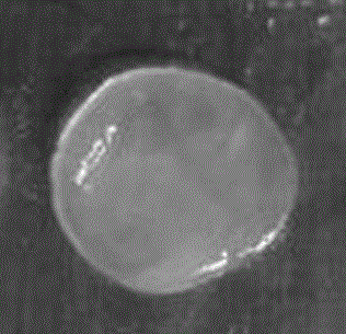
  • Preparation method of tissue engineering cornea
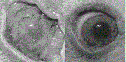
  • Preparation method of tissue engineering cornea
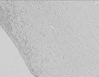
  • Preparation method of tissue engineering cornea

Examples


preparation example Construction

[0039] The preparation method of the tissue-engineered cornea mainly includes animal-derived cornea pretreatment, separation of each layer of the cornea, decellularization, virus inactivation treatment, preparation of cell suspension and construction of the tissue-engineered cornea. The specific preparation process is as follows:

[0040] Step 1, animal-derived corneal pretreatment:

[0041] After the animal-derived cornea is taken, soak it in deionized water at 2-30 ℃ for 8-24 hours with added antibiotics. The natural cornea will swell due to water absorption, and its thickness can swell to 2-5 times the thickness. After the corneal epithelium The cell layer will fall off due to the swelling of the Descemet's membrane. At the same time, the use of antibiotics can effectively remove the bacteria in the corneal tissue and obtain sterile materials.

[0042] The antibiotic described in this step is one or a mixture of two or more of penicillins, cephalosporins, streptomycin, gent...

Embodiment 1

[0065] Example 1. Preparation of tissue-engineered cornea of ​​porcine corneal scaffold composite cells.

[0066] Porcine corneas were collected and soaked in sterile deionized water mixed with gentamicin and streptomycin for 8 hours. The thickness of the pig cornea was swelled to twice the original thickness due to water absorption, and the epithelial cell layer fell off at the same time. Then place the above cornea on a microscopic table, cut the cornea into three layers with a corneal cutter, and the cut thickness of the Bowman's layer, stroma and Bowman's layer accounted for 5%, 90% and 5% of the total thickness respectively . The above tissue slices were then soaked repeatedly in 3M sodium chloride high-salt solution and deionized water for 15 cycles to remove cells, and at the same time irradiated with 15kGy for virus inactivation treatment. The tissue pieces were then placed in 85% glycerol and dehydrated at 25°C for later use.

[0067] At the same time, human corneal...

Embodiment 2

[0069] Example 2. Preparation of tissue-engineered cornea with bovine corneal scaffold composite cells.

[0070] The bovine cornea was collected and soaked in sterile deionized water added with penicillin for 24 hours. The thickness of the bovine cornea was swelled to 5 times of the initial thickness due to water absorption, and the epithelial cell layer fell off at the same time. Then the cornea was stratified by femtosecond laser, and it was divided into anterior descemet slices, stroma tissue slices and descemet tissue slices, whose thickness accounted for 10%, 80% and 10% of the total thickness. Soak the above tissue in 1M sodium hydroxide solution for 1 hour, wash it with deionized water, remove sodium hydroxide residue, remove cell structure, control the immunogenicity of the material, use 1% sodium hypochlorite virus inactivation treatment, 25kGy radiation Sterilize, and finally place in 95% glycerol at 30°C for dehydration until use.

[0071] At the same time, human c...


PUM

PropertyMeasurementUnit
Phaaaaaaaaaa
Ph valueaaaaaaaaaa

Abstract

The invention belongs to the technical field of tissue engineering and provides a preparation method of a tissue engineering cornea. A support material prepared from human corneal limbal cell compound animal source cornea is adopted, a bowman membrane, a substrate layer and a descemet membrane are separated and are coated with cell suspension prepared from human corneal limbal cells, and then crosslinking and bonding are carried out, so that construction of a cell-containing tissue engineering cornea is completed, and the tissue engineering cornea prepared by adopting the preparation method has the advantages of high tissue compatibility and industrialization; besides, the adopted cells are not subjected to in-vitro multiplication culture, cell characters are stable, differentiation is hardly caused, and the safety of the tissue engineering cornea is guaranteed.

Description

Technical field [0001] The present invention is an organizational engineering technology field, which involves a method of building corneal corneal. Background technique [0002] A cornea is one of the tissues that maintain the normal vision of the human body. The vision loss or blindness caused by the acute damage, perforation, ulcer, and lesions of the cornea, and even the eyeball removal, which has a great impact on the patient's normal life and psychological psychology. [0003] Nowadays, corneal blindness has become the second general blind disease in the world. The most effective method for treating corneal blindness is corneal transplantation. However, the lack of corneal sources, which causes patients to be treated in a timely manner and blind.According to statistics, about 4 million corneal blind patients in my country need corneal transplantation, but less than ten cases of patients have corneal transplantation.The lack of corneal supply sources restricts the treatment ...

Claims


Application Information

Patent Timeline
IPC IPC(8): A61L27/38A61L27/36
Inventor 张蓉
Owner SHAANXI BOYU REGENERATIVE MEDICINE CO LTD
Learn More