Anti-human NMP-22 monoclonal antibody and detection kit thereof
A technology of NMP-22 and monoclonal antibody, applied in the field of molecular immunology, can solve the problems of low sensitivity, time-consuming and labor-intensive kit operation, etc.
Active Publication Date: 2015-11-04
BEIJING PERGRANDE BIOTECH DEV
1 Cites 12 Cited by
AI-Extracted Technical Summary
Problems solved by technology
Human NMP-22 detection enzyme immunoassay kit (product number D1100E) manufactured by American Alere company occupies a large proportion in the market of majo...
Abstract
The present invention relates to an anti-human NMP-22 monoclonal antibody and a detection kit thereof, and particularly provides human nuclear matrix protein 22 (NMP-22) that is expressed by using a gene cloning recombinant technology, and an anti-human NMP-22 mouse monoclonal antibody is obtained by application of an immunological technology. The present invention further discloses a kit for specific detection of NMP-22 in a human sample. The kit comprises two specific anti-human NMP-22 monoclonal antibodies. The kit disclosed by the invention can be used for carrying out quantitative detection on the content of NMP-22 in a sample quickly, sensitively and specifically.
Application Domain
Immunoglobulins against animals/humansGenetic engineering +4
Technology Topic
Immunologic TechniqueMurine monoclonal antibody +6
Image
Examples
- Experimental program(3)
- Effect test(4)
Example Embodiment
[0050] Example 1. Preparation and purification of recombinant human NMP-22 protein
[0051] 1. Perform the operation according to the instructions of the EASYspin Whole Blood RNA Rapid Extraction Kit (Cat. No. RN06) produced by Beijing Bomed Gene Technology Co., Ltd. to extract whole blood RNA.
[0052] 2. Perform the operation according to the instructions of the Transcript First-strand cDNA Sythesis SuperMix Kit (Cat. No. AT301-02) produced by Beijing Quanshijin Biotechnology Co., Ltd., using the following reaction system:
[0053] cDNA first strand synthesis reaction system
[0054]
[0055] After incubating at 42°C for 30 minutes, the reverse transcriptase was inactivated at 85°C for 5 minutes to terminate the reaction.
[0056] Among them, Oligo (DT) 18, 2× reaction mixture solution, and enzyme mixture all come from the above kit, and the RNA comes from the whole blood RNA extracted in step 1.
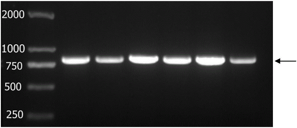
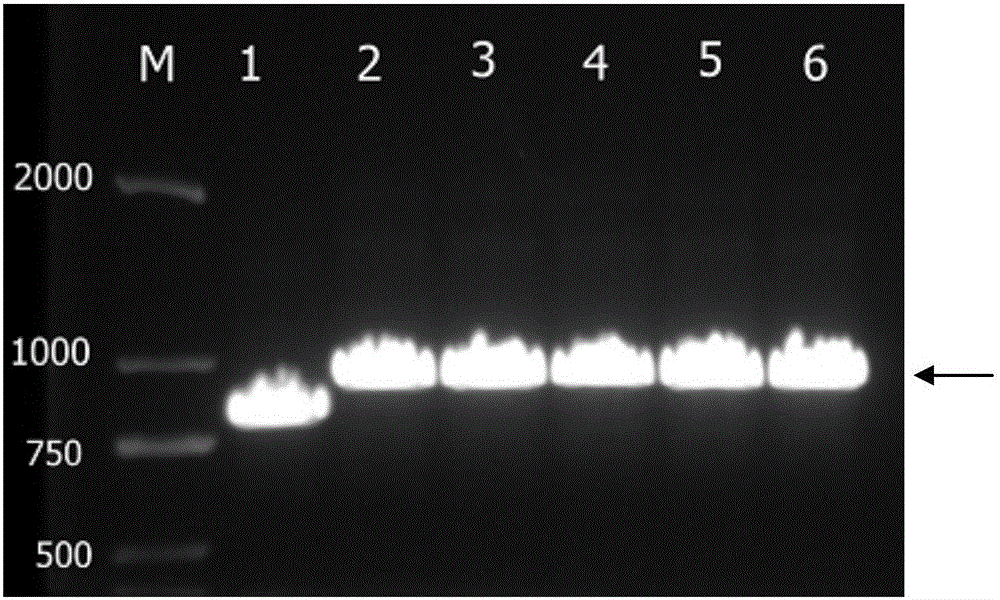
[0057] 3. Perform PCR reaction to amplify the target gene of NMP-22 according to the following reaction system and reaction conditions:
[0058] (1) Preparation of PCR reaction system
[0059]
[0060] (2) PCR reaction conditions
[0061]
[0062] The 2× reaction mixture solution is 2×TransTaq-T PCR SuperMix (Cat. No. AS122) produced by Beijing Quanshijin Biotechnology Co., Ltd. The main components include DNA polymerase, dNTP and reaction buffer. NMP-22-upstream primer and NMP-22-downstream primer are based on the gene coding region sequence of human NuMA registered in GenBank (accession number BC043499.1), and partial gene fragment 140-1057bp (SEQ ID No.2) is selected for cloning and recombination . Use primer-primer 5.0 primer design software for primer design, and add BamHI and XhoI restriction enzyme sites to the 5'ends of the upstream primer and downstream primer respectively, and send them to Beijing Bomaide Gene Technology Co., Ltd. for synthesis. The primer sequence as follows,
[0063] NMP-22 upstream primer (5'-3'): GGATCCATGACACTCCACGCCAC (SEQ ID NO. 3);
[0064] NMP-22 downstream primer (5'-3'): CTCGAGTTACTTCTCTGTCTTCAGGTC (SEQ ID NO. 4).
[0065] 4. Carry out agarose gel electrophoresis on PCR amplification reaction products, see the electrophoresis results figure 1. The amplified DNA band size is between 750-1000bp, which is consistent with the 918bp position of the NMP-22 target gene fragment. Use the agarose gel recovery kit (Cat. No. H41118) produced by Beijing Quanshijin Biotechnology Co., Ltd. to perform the operation according to the manufacturer's instructions to recover the PCR amplification products.
[0066] 5. Using the Beijing Quanshijin Biotechnology Co., Ltd. T vector ligation kit (Cat. No. H20724), according to the instruction manual, the DNA recovery product and pEASY-T1Simple vector were ligated at 25°C for 15 minutes. Add 10μl of the ligation product to 50μl of E. coli DH5α competent cells, incubate on ice for 30min, heat stress at 42°C for 90s, immediately place on ice for 2min, add 500μl of LB liquid medium, and culture with shaking at 200-250rpm at 37°C After 45 minutes, spread the bacterial solution on a screening culture plate with ampicillin resistance. After the plate is placed in the forward direction for 10 minutes, dry the residual bacterial solution, invert the plate, and incubate at 37°C overnight.
[0067] 6. Randomly pick the 6 single colonies in step 5 and place them in 3ml LB medium, and conduct overnight expansion culture at 37°C. Using bacterial liquid as cDNA template, refer to step 3 for PCR identification of bacterial liquid, and agarose gel electrophoresis, the results are shown figure 2. From figure 2 It can be seen that the positions of amplified DNA with clones No. 1 to No. 6 as templates are between 750-1000 bp, which is consistent with the size and position of the target gene NMP-22 (918 bp). The clones No. 1, No. 2 and No. 3 were selected and sent to Beijing Bomed Gene Technology Co., Ltd. for DNA sequencing. The feedback sequencing report was compared with the gene coding region sequence of human NuMA registered in GenBank (accession number BC043499.1). Among them, the sequence of the sequenced gene of clone No. 3 is completely consistent with the target gene sequence. See the sequence comparison result image 3. A small amount of plasmid DNA extraction kit (Cat. No. H41105) produced by Beijing Quanshijin Biotechnology Co., Ltd. was used to perform operations according to the kit instructions to extract plasmid DNA from the NMP-22-pEASY-T1Simple No. 3 clone. The extracted DNA was verified by double enzyme digestion with restriction enzymes BamHI (product number #R0136V) and XhoI (product number #R0146V) produced by New England Biotechnology (Beijing) Co., Ltd. The digested products were agarose gel electrophoresis and electrophoresis. See the result Figure 4. The position indicated by the arrow is the fragment released by restriction digestion, the size is between 750-1000bp, which is consistent with the size of the target gene NMP-22 (918bp).
[0068] 7. Use the restriction endonucleases BamHI (article number #R0136V) and XhoI (article number #R0146V) produced by New England Biotechnology (Beijing) Co., Ltd., according to the manufacturer's instructions, to compare the NMP-22-pEASY-T1 plasmid and prokaryotic Expression vector pET-28a plasmid (see the vector map Figure 5 , The position marked in the figure is the insertion position of the target gene NMP-22) Double enzyme digestion to make it have complementary sticky ends. Use the agarose gel recovery kit (Cat. No. H41118) produced by Beijing Quanshijin Biotechnology Co., Ltd., and perform operations according to the manufacturer's instructions to recover the target gene product with complementary sticky ends and the digested vector fragments separately. Using the T4DNA Ligase kit (Cat. No. D2011A) produced by Dalian TaKaRa Company, according to the manufacturer's instructions, the target gene product with complementary sticky ends and the digested vector fragment were ligated in a 16°C water bath for 12 hours. Add all the ligation products to 50μl of E. coli DH5α competent cells, incubate on ice for 30min; after heat stress at 42°C for 90s, immediately place on ice for 2min; add 500μl of LB liquid medium and culture at 37°C with 200-250rpm shaking for 45min; Spread the bacterial solution on a screening culture plate with ampicillin resistance. After the plate is placed in the forward direction for 10 minutes, dry the residual bacterial solution; invert the plate and incubate overnight at 37°C. Randomly pick 2 single colonies of pET-28a-NMP-22 in 3ml LB medium, and expand overnight at 37°C. A small amount of plasmid DNA extraction kit (Cat. No. H41105) produced by Beijing Quanshijin Biotechnology Co., Ltd. was used to perform operations according to the kit instruction manual to extract plasmid DNA from pET-28a-NMP-22. The extracted DNA was verified by double enzyme digestion with restriction enzymes BamHI (product number #R0136V) and XhoI (product number #R0146V) produced by New England Biotechnology (Beijing) Co., Ltd. The digested products were agarose gel electrophoresis and electrophoresis. See the result Image 6. The position indicated by the arrow is the digested fragment, the size is between 750-1000bp, which is consistent with the size of the target gene NMP-22 (918bp).
[0069] 8. For the prokaryotic expression plasmid pET-28-NMP-22 constructed in step 7 to transform BL21(DE3) competent cells, pick a single colony into 5ml LB liquid medium (containing kanamycin) at 220 rpm, 37 Incubate overnight at ℃. The overnight culture bacteria were transferred to fresh LB liquid medium at a ratio of 1:100, 220 rpm, 37 ℃ culture, when the bacterial concentration OD600 ≈ 0.6, add IPTG to start induction (IPTG final concentration is 0.25 mM); 28 ℃ 220 rpm induction 4h Receiving bacteria; ultrasonically disrupting the bacteria, ultrasonic conditions: every 3 seconds interval of 7 seconds, ultrasonic 5min, output power 50%. Centrifuge at 9500 rpm, 4°C for 20 min, separate the supernatant and precipitate, and identify them by SDS-PAGE polyacrylamide gel electrophoresis ( Figure 7 ), in which lane 1 is the negative whole bacteria induced without IPTG, lane 2 is the sedimentation lane of the ultrasonically induced bacteria, and lane 3 is the supernatant of the ultrasonically induced bacteria. The position indicated by the arrow is the expressed target protein, the size is between 30kd-40kd, which is consistent with the size (34kd) of the target protein NMP-22, and most of the protein is soluble in the supernatant form.
Example Embodiment
[0071] Example 2. Preparation of anti-human NMP-22 mouse monoclonal antibody
[0072] 1. Select 5 female Balb/c mice that are 6 weeks old and weigh about 20 g. The recombinant protein prepared in Example 1 was immunized. For the first immunization, take 100μg of protein plus 0.01M PBS to 500μl, emulsify it with an equal volume of Freund's complete adjuvant, and perform subcutaneous injections after sufficient emulsification. The second and third immunization doses were performed on the 14th and 35th days, respectively, with the same dose as the first immunization, and emulsified with Freund's incomplete adjuvant. After sufficient emulsification, multiple subcutaneous injections were performed. One week after the three immunizations, tail vein blood was taken from the mouse, and the recombinant antigen was coated with the indirect Elisa method for serum titer detection. The mouse with the highest titer (B mouse, see Picture 9 )'S spleen undergoes cell fusion. Three days before fusion, the immunization was boosted, and a dose of 20 μg recombinant protein was diluted to 100 μl 0.01M PBS and injected into the tail vein of mice; 3 days later, spleen was taken for cell fusion.
[0073] 2. Take the mice that were boosted 3 days in advance, pull their necks to death, and take the spleen aseptically. Wash once with 10ml of RMPI-1640 (1802-E) serum-free culture medium produced by Beijing Newtech Biotechnology Co., Ltd. The spleen is crushed and passed through a 200-mesh cell sieve. The spleen cells were transferred to a 10ml centrifuge tube and centrifuged at 800rpm for 10min. Wash the cells twice with 10ml culture medium, culture and resuspend the cells with 5ml serum-free, and count the cells under the microscope, a total of about 1×10 8 A suspension of splenic lymphocytes for use. Harvest the mouse myeloma SP2/0 cells in the logarithmic growth phase, wash them twice with 10ml serum-free culture medium, resuspend the cells in 5ml serum-free culture, and count the cells under the microscope, a total of about 5×10 7 A SP2/0 cell suspension is reserved.
[0074] 3. Cell fusion: mix myeloma cells and spleen cells in a ratio of 1:2, wash once with serum-free incomplete culture medium in a 50ml centrifuge tube, centrifuge, 1200rpm, 8min; discard the supernatant and use a pipette Absorb the remaining liquid so as not to affect the concentration of polyethylene glycol (PEG). Gently tap the bottom of the centrifuge tube to loosen the cell pellet slightly. Add 1ml 50% (0.5g/1ml) PEG polyethylene glycol (molecular weight 4000) solution pre-warmed at 37°C, and shake it slightly while adding. 37°C water bath for 90s. Add incomplete medium pre-warmed at 37°C to stop the effect of PEG, and add 1ml, 2ml, 3ml, 4ml, 5ml and 6ml every 2min. Centrifuge, 800rpm, 6min, discard the supernatant, and resuspend the cell suspension with 20% (v/v) special grade fetal bovine serum and 1×HAT medium additive Hybri-Max (Sigma Catalog No. H0262). In a 96-well cell culture plate, add 200μl to each well. Place the culture plate in a 37°C, 5% CO2 incubator for culture.
[0075] 4. After 10 days of cell fusion, observe under a microscope, when the hybridoma cell clones cover 1/5 of the hole bottom area, the indirect Elisa method can be used to detect the culture supernatant and screen the secretion-resistant positive cell clones. Dilute the recombinant protein to 5μg/ml with 0.05M carbonate buffer, coat a 96-well microtiter plate with 100μl/well, overnight at 4°C, pour out the liquid in the microplate well, add PBST, and repeat washing three times; 200μl/well of insulation solution (0.5% BSA, diluted with PBST) was sealed, placed at 37°C for 1 hour, and patted dry for later use. Add 100μl of cell culture supernatant, positive control with 1:1000 fusion mouse immune serum diluted with incubation solution, negative control with SP2/0 culture supernatant, blank as washing solution, and let stand at 37°C for 45 minutes. Wash the plate with 200μl/well PBST and repeat the washing three times. Add 100μl/well of HRP-labeled goat anti-mouse secondary antibody diluted 1:5000, and let stand at 37°C for 45 minutes. Wash the plate with 200μl/well PBST and repeat the washing three times. Add 100μl/well of the substrate reaction solution and react at 37°C in the dark for 10 minutes. Join H 2 SO 4 (2mol/L) 50μl/well, stop the reaction. The microplate reader detects the absorbance value at 450nm, compares the OD value of the positive control, and selects the cloning well with the OD value of the positive control equal to or greater for cloning.
[0076] 5. The positive clones were screened, and the hybridomas were cloned by the limiting dilution method. The hybridoma cells to be cloned were used with 20% (v/v) special grade fetal bovine serum and 1×HT medium additive (Sigma H0137) Select the culture medium to gently blow dry from the culture hole, count the cells, and adjust the cells to 5 cells/ml; add 200μl of diluted cells to each hole, culture in a 37°C, 5% CO2 incubator. The formation of cell clones can be seen in 10 days. Take the culture supernatant of the monoclonal cell wells for indirect Elisa detection, and clone the strongest positive monoclonal again until the positive rate of the monoclonal cells reaches 100%, then the strain can be expanded and cultivated. Perform cell cryopreservation. After 3 rounds of cloning, a total of 13 cell lines were retained, with clone numbers 7H3, 5E8, 1E12, 1D12, 4E4, 11A10, 8A11, 5D1, 3H7, 6A2, 13H5, 11F9, and 13A4.
[0077] 6. Inject 500 μl/head of paraffin oil into Balb/c mice by intraperitoneal injection. After 7 days, press the 13 hybridoma cells obtained in step 5 to 1×10 6 Intraperitoneal injection was performed at the concentration of cells, and ascites was collected 10 days later. Use Protein G Sepharose 4Fast Flow manufactured by GE Healthcare China to purify ascites by affinity chromatography according to the product manual.
Example Embodiment
[0080] Example 3: Preparation of human NMP-22 chemiluminescence kit
[0081] 1. Prepare a microtiter plate coated with NMP-22 primary antibody:
[0082] a) Coating: using a 0.05M carbonate buffer with a pH of 9.6 and an appropriate concentration of the mouse monoclonal antibody clone number 5E8 prepared in Example 2 to prepare coating solutions of different concentrations. Choose the coating concentration of 5μg/ml, 2.5μg/ml, 1.25μg/ml, 0.625μg/ml, and load the coating solution on the microplate with 120μl/well, and coat at 4℃ for 12h;
[0083] Standard formula of carbonate buffer:
[0084]
[0085] b) Washing the plate: Dilute the 20-fold concentrated lotion to 1 times the concentration, use the 1-fold concentration lotion to wash the plate twice; 20 times the concentrated lotion standard formula:
[0086]
[0087] c) Blocking: Load the blocking solution (disodium hydrogen phosphate 5.8g, sodium dihydrogen phosphate 0.593g, sodium chloride 8.0g, goat serum 100ml, Proclin-3000.5ml, purified water to a constant volume of 1000ml) on the microplate , Room temperature for 2 hours, spin dry, and dry overnight.
[0088] 2. Preparation of biotinylated antibody:
[0089] The Sulfo-NHS-LC-Biotinylation kit (Cat. No. 21435) manufactured by Thermo Fisher Scientific was used according to the kit instruction manual to label the monoclonal antibody clone number 11A10 prepared in Example 2.
[0090] 3. Use the checkerboard method (matrix method) to determine the optimal working concentration of the coated antibody and biotinylated antibody. Use microplates coated in step 1 with a concentration of 5μg/ml, 2.5μg/ml, 1.25μg/ml, 0.625μg/ml, and biotinylated antibody from step 2 (working concentrations are 1:8k, 1:16k, 1:32k and 1:64k dilution), streptavidin-labeled HRP (working concentration is 1:2.5k, 1:5k, 1:10k) for experiment. Determine that the final coating concentration is 5μg/ml (equivalent to 600ng/well of anti-human NMP-22 monoclonal antibody), the working concentration of biotinylated antibody is 1:8k, and the working concentration of streptavidin-labeled HRP is 1: 5k.
[0091] 4. Preparation of biotin-labeled antibody working solution:
[0092] The biotinylated monoclonal antibody prepared in step 2 was dissolved in the enzyme buffer at a ratio of 1:8000. The formula of enzyme buffer is: 5.8g/L disodium hydrogen phosphate, 0.593g/L sodium dihydrogen phosphate, 8.0g/L sodium chloride, 200ml/L calf serum and 0.5ml/L Proclin-300. The final concentration of the biotinylated monoclonal antibody is 12 mg/L.
[0093] 5. Preparation of sample diluent:
[0094] 1000ml sample diluent includes 30.0g sodium chloride, 25.0ml goat serum, 5.0ml Tween-20, 0.5ml Proclin-300.
[0095] 6. Chemiluminescence liquid A contains luminol and luminescence enhancer, and chemiluminescence liquid B contains peroxide and luminescence enhancer.
[0096] 7. Horseradish peroxidase labeled streptavidin working solution: 200mL/L calf serum, pH 7.4 phosphate buffer, 100ng/mL horseradish peroxidase labeled streptavidin and 0.5mL/L L Proclin 300.
[0097] 8. Assignment and preparation of calibrators:
[0098] Operate according to the instruction of the comparison kit, calibrate with the working calibrator, and test the high-value human sample of NMP-22 diluted with the sample diluent. Under the conditions of different laboratories and different operators, the independent measurement was repeated 20 times, and the OD value (light absorption value) of the 20 measurements was obtained. Select the human source sample with stable OD value as the product calibrator, and calculate the average value According to the standard curve equation of the working calibrator prepared by pure product, The corresponding OD value is brought into the above equation, and the corresponding concentration value is obtained, which is the target value of the product calibrator. Use sample diluent to prepare calibrators according to the assignment results: 100ng/ml, 50ng/ml, 10ng/ml, 2ng/ml, 1ng/ml, 0ng/ml.
[0099] 9. Preparation of quality control products:
[0100] Collect human urine samples with a high value for clinical testing of human NMP-22 (the urine can be any commercially available urine sample or collected from a clinical institution), and dilute the high value human urine with a sample diluent to a desired concentration range. High value quality control: 40ng/ml to 60ng/ml; low value quality control: 1.6ng/ml to 2.4ng/ml. Store at 2-8°C for later use.
[0101] 10. Assemble the above-mentioned reagents into a kit. If necessary, you can also incorporate tools such as instructions for use and a sealing film in the kit.
PUM


Description & Claims & Application Information
We can also present the details of the Description, Claims and Application information to help users get a comprehensive understanding of the technical details of the patent, such as background art, summary of invention, brief description of drawings, description of embodiments, and other original content. On the other hand, users can also determine the specific scope of protection of the technology through the list of claims; as well as understand the changes in the life cycle of the technology with the presentation of the patent timeline. Login to view more.