Method and device for detecting free epithelial cell organ sources in blood

A technology of epithelial cells and detection methods, applied in biochemical equipment and methods, biological testing, material inspection products, etc., to achieve the effects of simple structure, accurate early diagnosis, and easy operation

Active Publication Date: 2015-11-18
SHANGHAI JIAO TONG UNIV
View PDF1 Cites 12 Cited by
  • Summary
  • Abstract
  • Description
  • Claims
  • Application Information

AI Technical Summary

Problems solved by technology

[0006] The present invention provides a method and device for detecting the source of free epithelial cells in blood to sol

Method used


Image

  • Method and device for detecting free epithelial cell organ sources in blood
  • Method and device for detecting free epithelial cell organ sources in blood
  • Method and device for detecting free epithelial cell organ sources in blood

Examples


Embodiment 1

[0042] Example 1 Detection device for rare cell proteome in blood

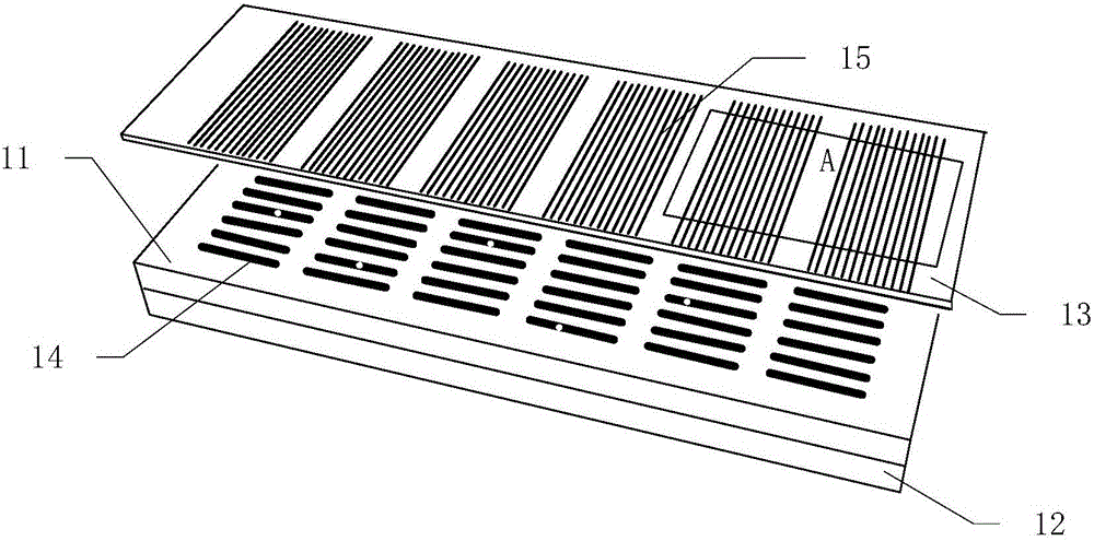
[0043] Such as figure 1 As shown, the device for detecting rare cell protein groups in blood according to the present invention includes a microgroove chip 11 , a magnet 12 and an antibody-loaded glass slide 13 .

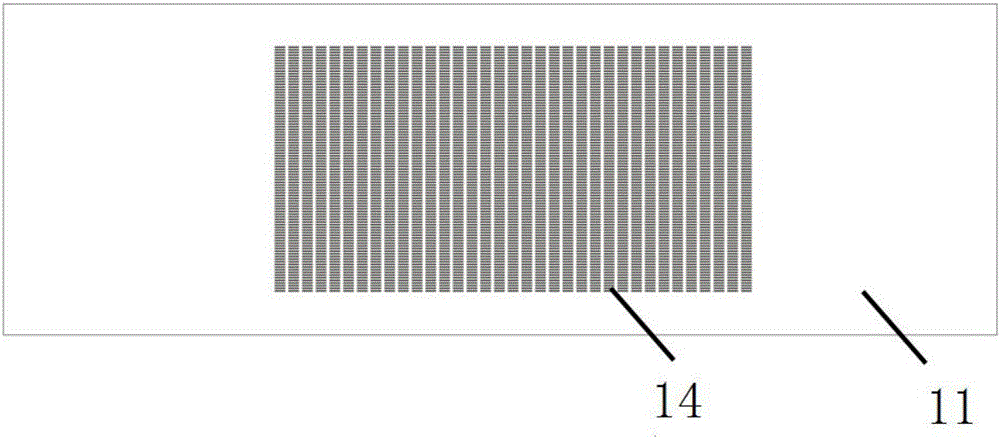
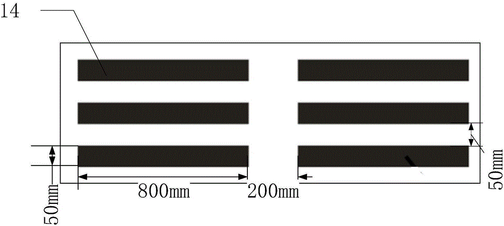
[0044] Such as figure 2 As shown, the microgroove chip 11 is provided with 3220 (35×92) microgrooves 14 with a volume of 1 nanoliter arranged in a matrix. Such as image 3 As shown, the cross-section of a single microgroove 14 is rectangular, with a length of 800 microns, a width of 50 microns, and a depth of 25 microns. During fabrication, a mask is first printed on a film substrate, a photoresist template is obtained through a photolithography process, and then a chip made of PDMS, that is, a microgroove chip 11 , is fabricated by using the photoresist template as a template.

[0045] Such as figure 1 As shown, the antibody-loaded glass slide 13 is loaded with an antibody microarray 15 , and...

Embodiment 2

[0049] Example 2 Detection of multiple organ-specific protein markers in circulating epithelial cells in human peripheral blood

[0050] In order to identify the organ source of the epithelial cells isolated from blood, a set of 8 organ-specific protein markers was designed in this example for identification.

[0051] Anti-TTF-1 (thyroid transcription factor 1), anti-GCDFP-15 (grosscystic disease fluid protein 15), anti-CDX2, anti-CK7, anti-CK20, anti-SMAD4, anti-CEA, anti - CD45, anti-CD105 (or anti-CD146) and single-stranded polynucleotide M are passed into different microchannels 19 of the antibody-loaded chip described in Example 1, respectively. Among them, anti-TTF-1 (thyroid transcription factor 1), anti-GCDFP-15 (grosscystic disease fluid protein 15), anti-CDX2, anti-CK7, anti-CK20, anti-SMAD4 and anti-CEA are used to specifically bind protein markers with organ specificity Anti-CD45 is used for specifically binding leukocyte markers, anti-CD105 (or anti-CD146) is use...

Embodiment 3

[0054] Example 3 Identification of Organ Source of Circulating Epithelial Cells

[0055] Adopt the method for embodiment 2 to have lung (100 examples), stomach (30 examples), colon (30 examples), ovary (10 examples), pancreas (10 examples) and breast ( 20 cases) samples from patients with malignant lesions were detected and organ source identification.

[0056] Cluster analysis was performed on the results of protein marker detection, combined with the existing biological knowledge about organ-specific markers and the location of malignant lesions in patients, the following protein-based identification method for epithelial cell organ localization was obtained.

[0057] First, remove the data of CD45+ cells (leukocytes) and CD105+ (or CD146+) cells (endothelial cells) in the test results;

[0058] Then, according to the rules in Table 1, the results of epithelial cell origin were obtained.

[0059] Table I

[0060]


PUM


Abstract

The invention relates to a method and a device for detecting free epithelial cell organ sources in blood. The method includes the following steps: capturing epithelial cells from a blood sample; cracking the epithelial cells to release protein therein; specifically recognizing at least four protein markers with organ specificity; acquiring the organ sources of the epithelial cells by detecting expression quantity of the protein markers. The device comprises a microgroove chip and an antibody loading glass piece, 100-10000 microgrooves are formed in the microgroove chip and used for containing the cells, the antibody loading glass piece is used for loading an antibody micro-array which comprises at least four mutually-independent antibody strips, and the microgrooves and the antibody micro-array are arranged in a matched manner to enable at least one part of each antibody strip to be contained at corresponding positions of the microgrooves. The problem of detecting the epithelial cell organ sources in blood is solved by quickly and effectively detecting multiple proteins in single cells.

Description

technical field [0001] The invention belongs to a method for detecting the organ source of epithelial cells, in particular to a method and a device for detecting the organ source of free epithelial cells in blood. Background technique [0002] Various major non-communicable diseases (such as tumors, cardiovascular and cerebrovascular diseases, diabetes, etc.) have entered a state of high growth in China, and the aging population and environmental pollution have further exacerbated this trend. Many major diseases currently lack effective treatment methods and the cost of treatment is high. While causing serious damage to individual health, it also imposes a heavy economic burden on families and society. Preventing the occurrence of these major diseases and their early detection and early intervention are effective ways to solve this problem. The so-called early detection means that the clues of major diseases can be found before the patients have obvious symptoms or before t...

Claims


Application Information

Patent Timeline
IPC IPC(8): C12Q1/04G01N33/68
Inventor 施奇惠邓宇亮王智华
Owner SHANGHAI JIAO TONG UNIV
Why Eureka
Try Eureka