MIP (macrophage inflammatory protein) 3alpha-Fc fusion protein and application thereof
A fusion protein and inflammatory protein technology, applied in the field of genetic engineering, can solve the problems of short half-life of MIP-3α protein and limited clinical application value, and achieve the effects of long-acting plasma half-life, growth inhibition, good biological activity and stability
- Summary
- Abstract
- Description
- Claims
- Application Information
AI Technical Summary
Problems solved by technology
Method used
Image
Examples
Embodiment 1MI
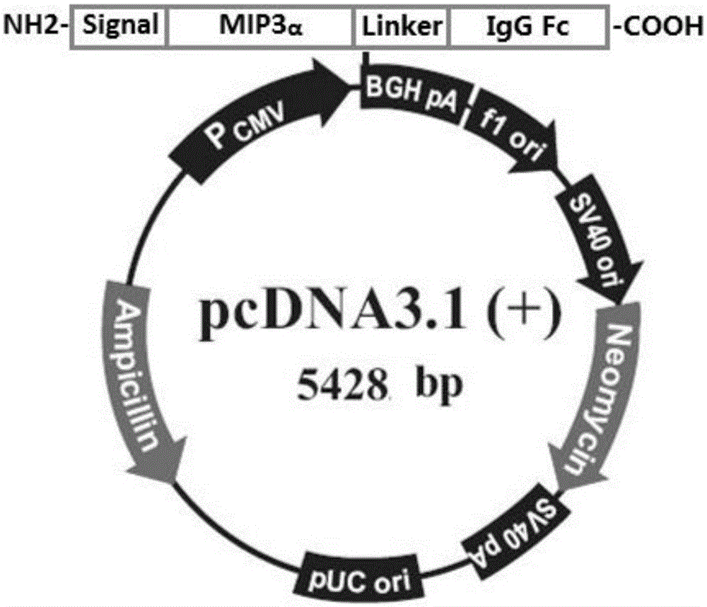
[0026] Example 1 Construction of MIP3α-Fc Fusion Protein Recombinant Expression Plasmid
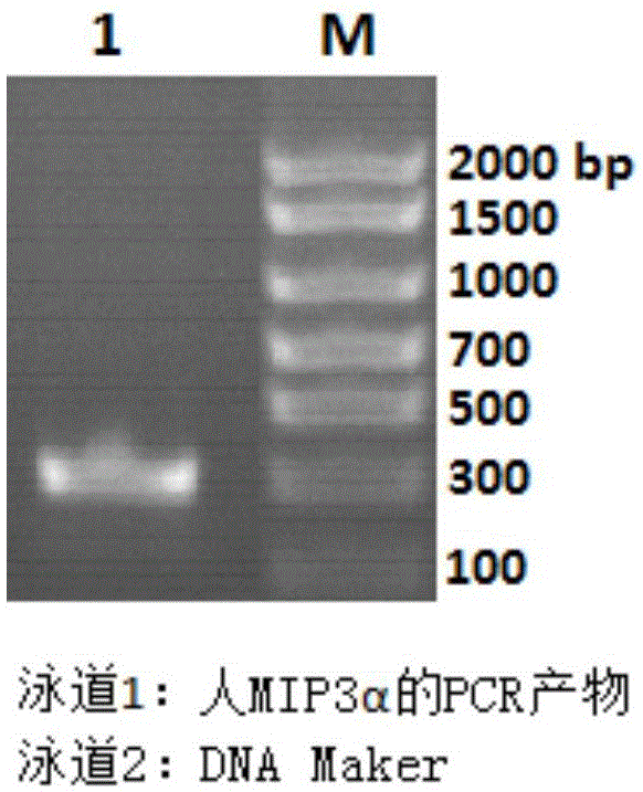
[0027] 1. Construction of human MIP3α-Fc fusion protein recombinant expression plasmid
[0028] The human MIP3α gene (GeneBank: NM_001130046.1) was amplified using the cDNA clone (purchased from Origene) as a template, and the primer sequences used were as follows (including Kozak sequence, cloning site, and protected bases)
[0029] Upstream primer: 5'-ATATCCTTAAGCGGCCGCCGCCACCATGTGCTGTACCAAG-3'
[0030] Downstream primer: 5'-ATATGGGATCCATGTTCTTGACTTTTTACTG-3'
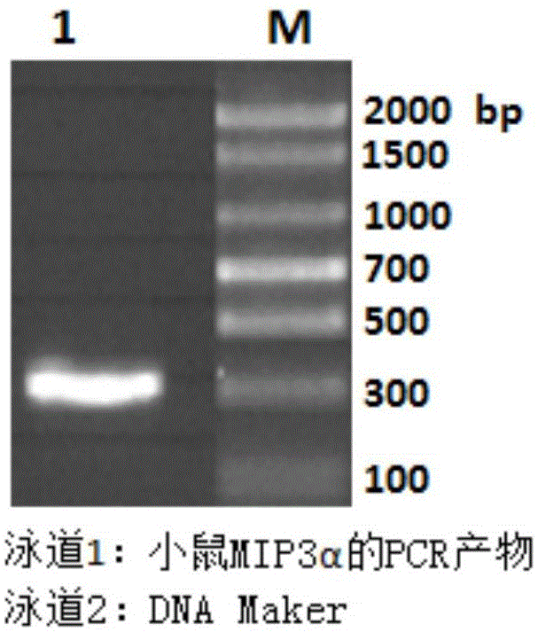
[0031] Use 50ul of PCR reaction system, add 1ul of 20mM primers; add 1ul of 10mM dNTP; add 1ul of pfuDNA polymerase, 2.5U / ul. The reaction conditions were 30 cycles of 95°C for 30 seconds, 55°C for 30 seconds, and 72°C for 45 seconds. The PCR product was analyzed by 1.8% agarose gel electrophoresis, which was in line with the expectation (320bp).
[0032] The obtained PCR product was recovered by gel and digested with AfLII a...
Embodiment 2M
[0039] Example 2 Expression and purification of MIP3α-Fc fusion protein
[0040] 1. Transient expression of MIP3α-Fc fusion protein in HEK293 cells
[0041] HEK293 cells in the logarithmic growth phase were digested with trypsin and diluted to 6×10 5 / mL density, inoculate into 15cm petri dish, add 20ml serum-free medium to each petri dish, and culture for about 24 hours for transfection. The recombinant plasmids were transfected into HEK293 cells using Lipofectamine2000 transfection reagent (Invitrogen), and the plasmids used were extracted by an endotoxin-free plasmid extraction kit (Sigma). Transfected cells were cultured in a 5% carbon dioxide incubator for 5 days and the supernatant was obtained. The fusion protein yield in the supernatant was determined by enzyme-linked immunosorbent assay (ELISA).
[0042]2. Stable high expression of MIP3α-Fc fusion protein in CHO cells
[0043] Dilute CHO cells in log phase to 4x10 6 After / ml, add 0.5ml to the electric shock cup,...
Embodiment 3MI
[0046] Example 3 Determination of plasma half-life of MIP3α-Fc fusion protein
[0047] Four 10-week-old C57BL / 6 mice were injected with mouse MIP3α-Fc fusion protein into the tail vein, 0.1 mg per mouse, and blood was collected alternately at the following time points: 30 minutes, 2 hours, 4 hours, 12 hours, 24 hours, 48 hours, 72 hours, 96 hours. Blood samples were used to detect the concentration of mouse MIP3α-Fc fusion protein by specific ELISA. The results showed that the half-life of the MIP3α-Fc fusion protein was as long as 30 hours. The result is as Figure 6 shown.
PUM
 Abstract
Description
Claims
Application Information
 - R&D
- Intellectual Property
- Life Sciences
- Materials
- Tech Scout
- Unparalleled Data Quality
- Higher Quality Content
- 60% Fewer Hallucinations
Browse by: Latest US Patents, China's latest patents, Technical Efficacy Thesaurus, Application Domain, Technology Topic, Popular Technical Reports.
© 2025 PatSnap. All rights reserved.Legal|Privacy policy|Modern Slavery Act Transparency Statement|Sitemap|About US| Contact US: help@patsnap.com


