Method capable of promoting migration of dendritic cells to lymph nodes and achieving multi-mode imaging simultaneously

A multi-mode imaging and dendritic cell technology, applied in the field of biological sciences, can solve the problems of low DC migration efficiency, ineffective pre-stimulation, adverse reactions, etc., and achieve improved living migration efficiency, good biocompatibility, and biological good compatibility

Active Publication Date: 2016-04-13
HUAZHONG UNIV OF SCI & TECH
View PDF2 Cites 8 Cited by
  • Summary
  • Abstract
  • Description
  • Claims
  • Application Information

AI Technical Summary

Problems solved by technology

Elucidating the migration mechanism of DC can guide the design of DC vaccine in the future to a certain extent, but the following problems exist in the pre-stimulation of inflammatory factors: 1) The migration rate of DC is still low (2×10 6 ), pre-stimulation basically does not work; 3) high doses of inflammatory factors in vivo administration h

Method used


Image

  • Method capable of promoting migration of dendritic cells to lymph nodes and achieving multi-mode imaging simultaneously
  • Method capable of promoting migration of dendritic cells to lymph nodes and achieving multi-mode imaging simultaneously
  • Method capable of promoting migration of dendritic cells to lymph nodes and achieving multi-mode imaging simultaneously

Examples


preparation example Construction

[0034] 1) Preparation of mDCs: The preparation of DCs is derived from bone marrow extraction and isolation. The preparation of mDCs is obtained by conventional culture methods, which may carry tumor antigens or not contain tumor antigens. DC can be extracted from bone marrow or DC2.4 cell line.

[0035] 2) mDCs carry polypeptide antigens and magnetic substances: the mDCs are incubated with a magnetic fluorescent nanoparticle at 37°C for 6 hours to complete.

[0036] The magnetic fluorescent nanometer particle is formed by organically combining oleate-iron oxide particles, two kinds of phospholipids, a near-infrared probe and an antigen fusion peptide with lipid binding ability through self-assembly.

[0037] The oleate-iron oxide particles are nanoparticles modified with oleic acid, which has been put into commercial production (see http: / / www.nanoeast.net / cxnmklfzxs.html, oleic acid-modified trioxide tetroxide Ferromagnetic nanoparticles (OAFe 3 o 4 , coprecipitation or hi...

Embodiment 1

[0057] The antigen fusion polypeptide constituting the magnetic fluorescent nanoparticle is composed of α-helical polypeptide (R4F), connecting sequence and antigenic peptide (AP) in series in the form of covalent bonds. The amino acid sequence of the α-helical polypeptide is: FAEKFKEAVKDYFAKFWD, the amino acid sequence of the connecting sequence is GSG, and the amino acid sequence of the antigenic peptide is: KVPRNQDWL. The amino acid sequence of the polypeptide is described in SEQ ID NO.1 in the sequence listing.

[0058] Prepare magnetic fluorescent nanoparticles, the steps are:

[0059] Preparation of magnetic fluorescent nanoparticles: Weigh 5 mg each of DMPC and hydroxypropyl methylcellulose ether (MHPC) and place them in EP tubes, then add 200 μl of chloroform to the EP tube for dissolution, and dissolve the dissolved DPMC and MHPC were transferred into a chloroform solution containing iron oxide (10 mg / mL) with a volume of about 400 μl, and methanol was added at a rat...

Embodiment 2

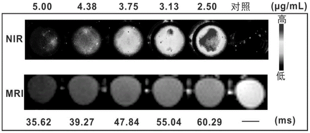
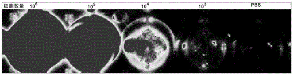
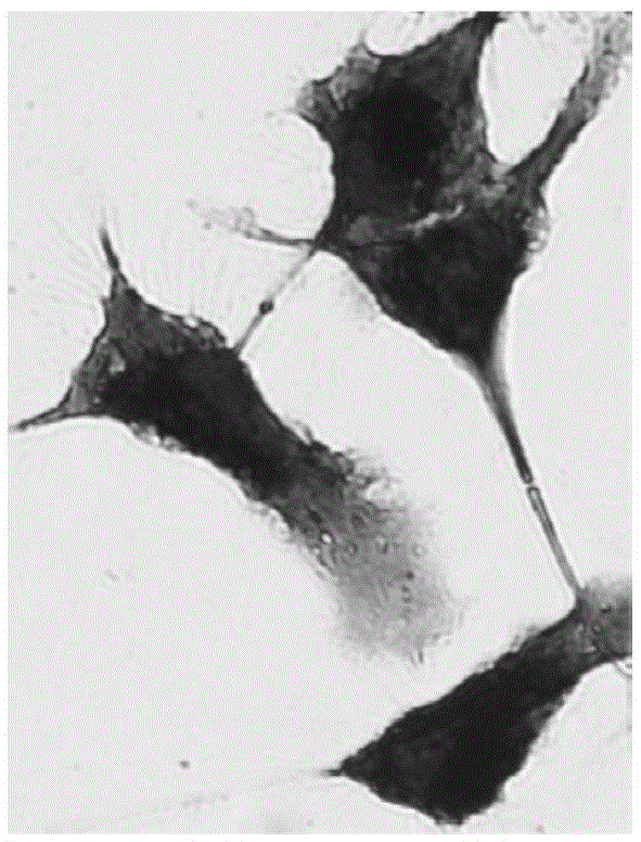
[0062] For the labeling ability of the magnetic fluorescent nanoparticles prepared in Example 1 to mature DC (mDC), see image 3 . image 3 It shows that the magnetic fluorescent nanoparticles can efficiently fluorescently label mDC, and at 50 μg / mL, it has no great influence on the morphology of mDC (within 20%), and the concentration selected in the experiment is 20 μg / mL. Figure 4 Cell survival experiments after co-incubation of mDCs with different concentrations of magnetic fluorescent nanoparticles are shown, and the results show that within the concentration range of 50 μg / mL, magnetic fluorescent nanoparticles have no significant effect on the growth of mDCs. mDCs that uptake magnetic fluorescent nanoparticles have fluorescent properties ( Figure 5 ), the fluorescence intensity increased with the increase of the number of cells. At the same time, Prussian blue staining proved that mDCs that ingested magnetic fluorescent nanoparticles contained a large amount of magn...


PUM


Abstract

The invention discloses a method capable of promoting migration of dendritic cells to lymph nodes and achieving multi-mode imaging simultaneously. The method is implemented by mDCs (mature dendritic cells) carrying magnetic fluorescent nanoparticles in an externally applied magnetic field. The magnetic fluorescent nanoparticles are formed by organically combining oleate-magnetic iron oxide particles, two phospholipids, near-infrared probes and an antigen fusion peptide with lipid binding capability through self-assembly. The method has the advantages that efficiency of DC living migration to a lymphoid tissue can be improved effectively, tumor prevention and treatment effects after DCs absorb antigen peptides are promoted, and multi-mode living imaging detection can be performed.

Description

technical field [0001] The invention belongs to the field of biological sciences, and in particular relates to a method for promoting the migration of dendritic cells to lymphoid tissue and performing multi-mode imaging monitoring on the migration process. Background technique [0002] DC vaccines are obtained by inducing DCs to be cultured in vitro using the patient's own monocytes, and then loaded with corresponding tumor antigens and stimulated to mature. To exert its therapeutic effect efficiently, mature DC (matureDC, mDC) must migrate to the patient's lymph node tissue and interact with naive T cells. A large amount of research evidence has shown that the in vivo immune response generated by DC is positively correlated with the migration efficiency of mature DC carrying relevant tumor antigens to secondary lymphoid tissues. Although recent pre-clinical experiments have shown that DC vaccines have good therapeutic effects, less than 2% of injected DCs reach secondary l...

Claims


Application Information

Patent Timeline
IPC IPC(8): A61K39/00A61K47/48A61K41/00A61K49/00A61K49/18A61K38/10A61K38/08A61P35/00
CPCA61K38/08A61K38/10A61K39/0011A61K41/00A61K49/0034A61K49/0093A61K49/1839A61K2039/5154A61K2039/585
Inventor 张智红金红林张宇戴艳锋
Owner HUAZHONG UNIV OF SCI & TECH
Why Eureka
Try Eureka