
Multifunctional fluorescent protein nanowire and nanowire-mediated immunoassay method

A fluorescent protein and green fluorescent protein technology, which is applied in the field of multifunctional fluorescent protein nanowires and its mediated immune analysis, can solve the problems of insufficient detection sensitivity and complicated preparation, and achieve improved sensitivity, simple preparation process, and non-specific adsorption. low effect

Inactive Publication Date: 2016-06-08
WUHAN INST OF VIROLOGY CHINESE ACADEMY OF SCI
View PDF6 Cites 8 Cited by
  • Summary
  • Abstract
  • Description
  • Claims
  • Application Information

AI Technical Summary

Problems solved by technology

[0010] In order to overcome the disadvantages of immunoassays in the prior art, especially the lack of detection sensitivity and complicated preparation of protein chips, the present invention provides a multifunctional fluorescent protein nanowire and its mediated immunoassay signal amplification method, using fluorescent molecules as signal molecules, Functional ligands are modified on the surface of fluorescent molecules located at both ends of multifunctional fluorescent protein nanowires as binding molecules, which can be used to improve the sensitivity of immunoassays

Method used


Image

  • Multifunctional fluorescent protein nanowire and nanowire-mediated immunoassay method
  • Multifunctional fluorescent protein nanowire and nanowire-mediated immunoassay method
  • Multifunctional fluorescent protein nanowire and nanowire-mediated immunoassay method

Examples


Embodiment 1

[0080] Example 1 Preparation of self-biotinylated fluorescent bifunctional protein

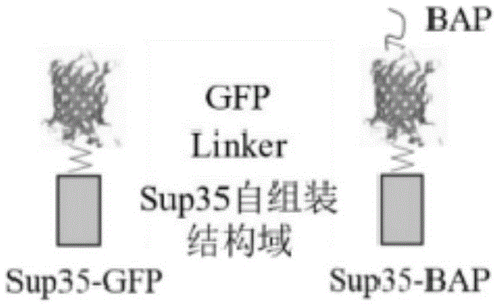
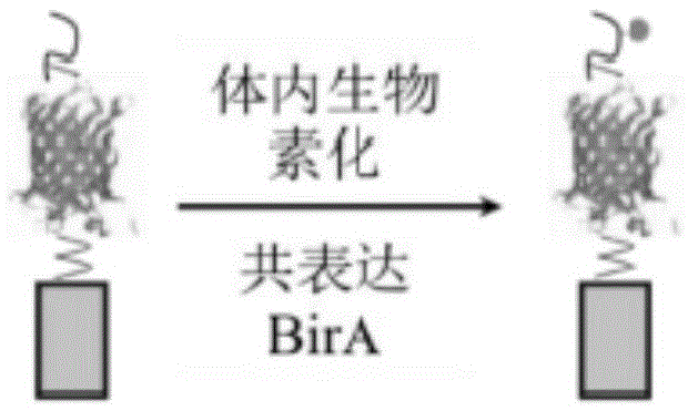
[0081] 1. Self-biotinylated fluorescent bifunctional protein cloning: through molecular cloning, the yeast prion protein self-assembly domain (1-61 amino acids of Sup35, referred to as Sup35) and green fluorescent protein (Green Fluorescent Protein, GFP) are flexibly connected to the polypeptide gene Fusion connection to form fusion protein Sup35-GFP (attached figure 1 left). A flexible polypeptide (LinkerPeptide) and a biotin-accepting peptide (BiotinAcceptedPeptide, BAP) are fused at the C-terminus of Sup35-GFP to form a fusion protein Sup35-GFP-BAP (Sup35-BAP for short, attached figure 1 right). The BAP tag in the fusion protein Sup35-BAP can be biotinylated under the action of Escherichia coli biotin ligase (Biotin-protein Ligase (EC6.3.4.15), BirA) (attached figure 2 ).

[0082] 2. Expression and purification of biotinylated fluorescent bifunctional protein: the gene of the fusion pr...

Embodiment 2

[0090] Example 2 Controllable Preparation of Multifunctional Fluorescent Protein Nanowires

[0091] 1. Preparation of Sup35-GFP seeds: After incubating the Sup35-GFP fusion protein at 4°C for one week, the long nanowires were broken into nanowire fragments by the shear force generated by ultrasound to prepare seeds (see attached Figure 5 Sup35-GFP seed electron microscope characterization result), the seed can quickly induce the fusion protein fused with the self-assembly domain to assemble at its end.

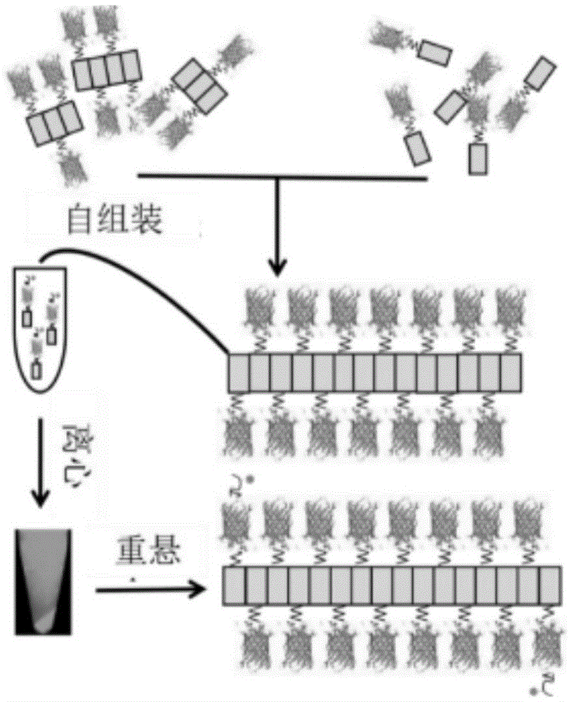
[0092] 2. Preparation of multifunctional fluorescent protein nanowires: mix prepared Sup35-GFP seeds with Sup35-GFP fusion protein at a certain molar ratio (1:1–1:16 ratio range), and incubate at 4°C for 8 hours for rapid seed-induced assembly. After the reaction, it can be found that there are obvious green fluorescent flocculent precipitates in the solution. Mix the reaction product with Sup35-BAP in equal proportions, centrifuge at low speed at 4°C, and collect the preci...

Embodiment 3

[0096] In a specific embodiment of the present invention, bFNPw can be used to prepare products detected by ELISA, such as sensors, chips, and kits. When in use, bFNPw described in the present invention is mixed with other existing commercial reagents, which can be used in various various forms of immunoassay.

[0097] Those skilled in the art know that the kit may also include a buffer, a washing solution, a diluent or a color developing agent, etc., which will not be repeated here.


PUM


Abstract

The invention provides a multifunctional fluorescent protein nanowire. The multifunctional fluorescent protein nanowire comprises a protein nanowire and fluorescent molecules connected to the outer side of the protein nanowire, wherein the protein nanowire is formed through self-assembly of wire forming protein. According to the multifunctional fluorescent protein nanowire, fluorescent molecules serve as signal molecules, the surfaces of the fluorescent molecules at the two ends of the multifunctional fluorescent protein nanowire are modified with function ligands to serve as binding molecules, and the multifunctional fluorescent protein nanowire can be used for improving the sensitivity of immunodetection, especially protein chip detection.

Description

technical field [0001] The invention relates to the field of immune analysis, in particular to a multifunctional fluorescent protein nanowire and an immune analysis method mediated by the same. Background technique [0002] Protein microarray analysis (Protein Microarray) technology, also known as protein chip technology, can be used for large-scale protein interaction screening, high-throughput target protein detection, various protein spectrum analysis, protein-nucleic acid interaction analysis, protein-small molecule ( drug screening) interaction analysis, etc. Its basic principle is similar to enzyme-linked immunosorbent assay (ELISA). A large number of different target proteins are immobilized on the surface of a solid phase carrier (glass slide, mica) in a specified order, and the position of the target protein is indicated by fluorescently labeled antibodies or interacting molecules. In order to obtain information about the interaction. Protein chip technology makes...

Claims


Application Information

Patent Timeline
Patent Type & Authority Applications(China)
IPC IPC(8): G01N33/68C12N15/62C12N15/63C07K19/00
CPCG01N33/6803C07K14/39C07K2319/60
Inventor 门冬张先恩周娟张治平
Owner WUHAN INST OF VIROLOGY CHINESE ACADEMY OF SCI
Learn More