Intelligent nanoparticle capable of specifically accelerating tumor cell apoptosis and monitoring curative effect by itself
A tumor cell apoptosis and self-monitoring technology, applied in the field of smart nanoparticles and their preparation, can solve the problems of accelerated tumor cell apoptosis, slow drug effect process, lack of curative effect monitoring, etc., achieves good tumor targeting, avoids Acid environment and enzymatic degradation, realizing the effect of timely monitoring
- Summary
- Abstract
- Description
- Claims
- Application Information
AI Technical Summary
Problems solved by technology
Method used
Image
Examples
Embodiment 1
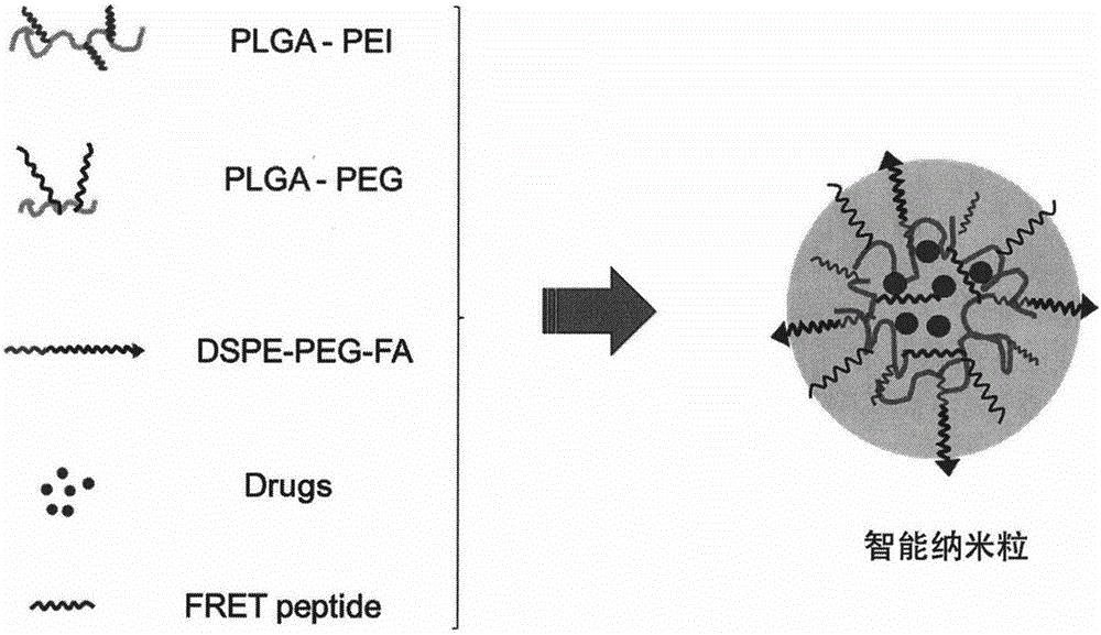
[0039] Example 1: Combining figure 1 , Synthesize smart nanoparticles that specifically accelerate tumor cell apoptosis and self-monitor curative effect
[0040] 1. Dissolve 1.6mg of hydrophobic antineoplastic drugs (CPT, PTX, DOX, CUR, EVO or SIL), 0.2mg of FRET-Pep, 5mg of PLGA-PEG and 4mg of PLGA-PEI in 2mL of DMSO, stir to dissolve completely;
[0041] 2. Dissolve 1mg DSPE-PEG-FA in 10mL ultrapure water;
[0042] 3. Add the mixture in the above step 1 dropwise to the mixture in the above step 2, and stir for 3-5 hours at room temperature in the dark;
[0043] After stirring, DMSO was removed by ultrafiltration (4000g, 15min), washed with ultrapure water and resuspended to obtain smart nanoparticles that specifically accelerate tumor cell apoptosis and self-monitoring of curative effect, and store at 4°C.
Embodiment 2
[0044] Example 2: Characterization of smart nanoparticles that specifically accelerate tumor cell apoptosis and self-monitor curative effect
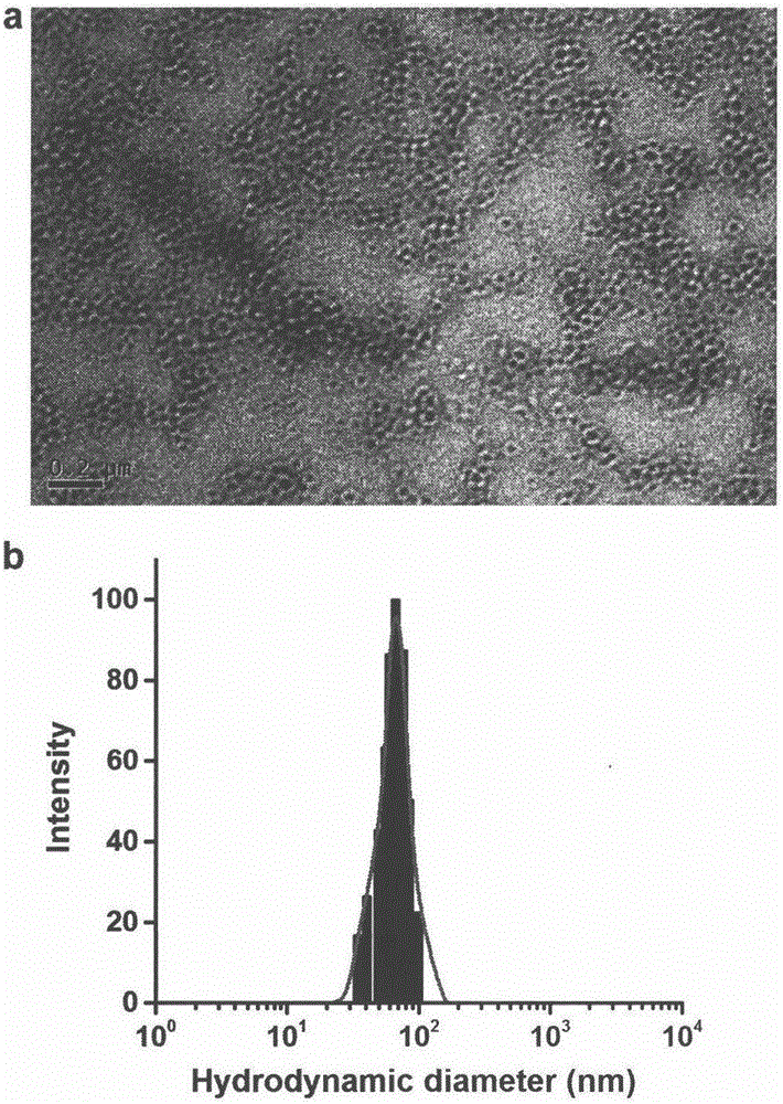
[0045] The morphology of intelligent nanoparticles that specifically accelerates tumor cell apoptosis and self-monitoring of curative effect is characterized by JEOL JEM-200CX transmission electron microscope (TEM), and the accelerating voltage is 200kV. The sample solution was dropped on the copper grid of the carbon support film, negatively stained with 2.0% (w / v) phosphotungstic acid solution, and observed under the electron microscope; the size and distribution were characterized by Mastersizer 2000 particle size analyzer dynamic light scattering (DLS). Depend on image 3 It can be seen that the nanoparticle has a spherical core-shell structure, and the nanoparticle has good dispersibility, uniform particle size, and an average particle size of 50-90 nm.
Embodiment 3
[0046] Example 3: Fluorescence response of FRET-Pep to caspase-3
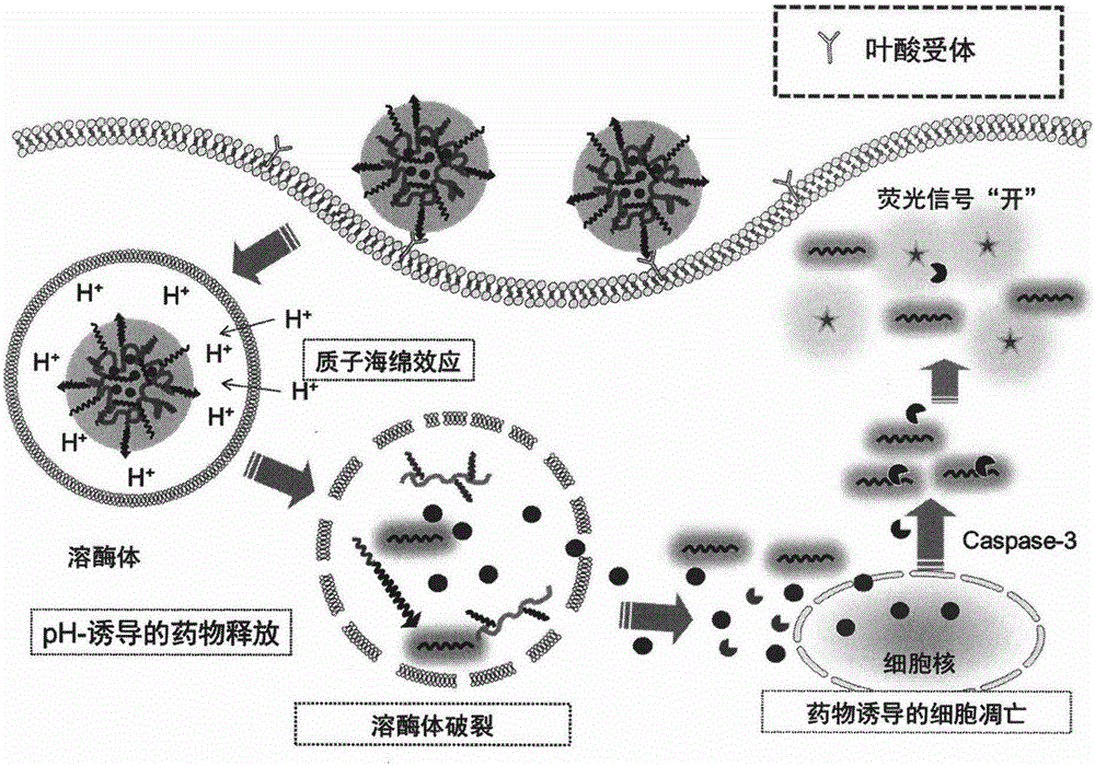
[0047] 100 μL of FRET-Pep was added to 900 μL of reaction buffer, pH 7.4, consisting of 50 mM 4-hydroxyethylpiperazineethanesulfonic acid (HEPES), 10 mM dithiothreitol (DTT), 100 mM sodium chloride ( NaCl), 1 mM ethylenediaminetetraacetic acid (EDTA), 0.1% w / v 3-[3-(cholamidopropyl)dimethylamino]propanesulfonic acid inner salt (CHAPS), 10% v / v glycerol (glycerol) . Add different concentrations of caspase-3, and react at 37°C for 1h. Depend on Figure 4 It can be seen that as the concentration of caspase-3 increases, the fluorescence of the polypeptide substrate gradually increases, indicating that the polypeptide substrate can specifically respond to caspase-3.
PUM
| Property | Measurement | Unit |
|---|---|---|
| The average particle size | aaaaa | aaaaa |
Abstract
Description
Claims
Application Information
 - R&D
- Intellectual Property
- Life Sciences
- Materials
- Tech Scout
- Unparalleled Data Quality
- Higher Quality Content
- 60% Fewer Hallucinations
Browse by: Latest US Patents, China's latest patents, Technical Efficacy Thesaurus, Application Domain, Technology Topic, Popular Technical Reports.
© 2025 PatSnap. All rights reserved.Legal|Privacy policy|Modern Slavery Act Transparency Statement|Sitemap|About US| Contact US: help@patsnap.com


