Separation culture method of laying duck small yellow follicle granular cells

A technology for granulosa cells and small yellow follicles, which is applied in the field of separation and culture of granulosa cells of small yolk follicles of laying ducks, can solve the problem that the separation and culture method of primary granulosa cells of laying duck follicles has not yet been established, and achieves high purity, good vitality, and operation. simple effect

Inactive Publication Date: 2016-08-24
ANIMAL SCI RES INST GUANGDONG ACADEMY OF AGRI SCI
View PDF2 Cites 6 Cited by
  • Summary
  • Abstract
  • Description
  • Claims
  • Application Information

AI Technical Summary

Problems solved by technology

The invention solves the problem of the method for separating and culturing primary granulosa cells of laying duck follicles that has not yet been established

Method used


Image

  • Separation culture method of laying duck small yellow follicle granular cells
  • Separation culture method of laying duck small yellow follicle granular cells
  • Separation culture method of laying duck small yellow follicle granular cells

Examples


Embodiment 1

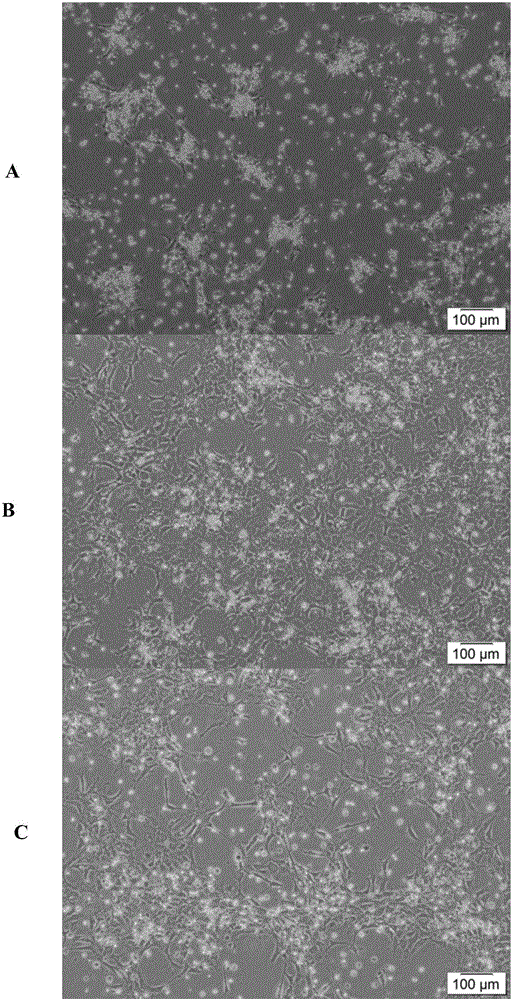
[0044] (1) Select 172-day-old egg-laying ducks, use wool scissors to cut off the feathers and fluff of the abdomen, bleed the neck veins, wash the wounds with sterilized water, and wash the whole body with Promethazine diluent (1:49) After 2 minutes, dry the body with a sterilized dry cloth, send it to the cell culture room, spray the whole body with 75% (v / v) alcohol, and transfer it to the ultra-clean bench.

[0045] (2) Separate the outer skin and muscle layer in turn, peel off the membrane tissue around the ovary, separate and cut off the ovary with blunt tweezers, take out the entire ovarian tissue and follicles, and put them into a beaker filled with PBS.

[0046] (3) After measuring and grading the diameter of the follicles with a vernier caliper, the small yellow follicles with a diameter of 3-5 mm were separated with ophthalmic scissors and transferred to a 12 cm sterilized petri dish filled with PBS.

[0047] (4) After peeling off the connective and vascular tissue of ...

Embodiment 2

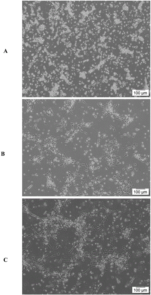
[0060] (1) Choose 185-day-old healthy laying ducks, kill them by bleeding from the jugular vein, wash the wound with sterilized distilled water, wash the whole body with bromogeramine diluent (1:49) for disinfection, dry it with a sterilized rag, put it in a tray, and transfer it to in the cell compartment.

[0061] (2) Spray the whole body of the duck with 75% (v / v) alcohol, and transfer it to the ultra-clean bench.

[0062] (3) Use surgical scissors to separate the skin layer, muscle layer and periovary membrane tissue, use forceps and surgical scissors to separate the complete ovary and follicles located on the left side of the duck body, and transfer them to a 50 mL sterilized beaker filled with PBS.

[0063] (4) Separate all the small yellow follicles with surgical scissors, measure the diameter of the follicles with a vernier caliper, and then grade them. Select the small yellow follicles with a diameter of 6-8 mm and transfer them to a 12 cm sterilized glass culture dis...


PUM

PropertyMeasurementUnit
diameteraaaaaaaaaa
diameteraaaaaaaaaa
diameteraaaaaaaaaa

Abstract

The invention discloses a separation culture method of laying duck small yellow follicle granular cells. The separation culture method includes following steps: selecting a laying duck, killing the laying duck by bloodletting a jugular vein, respectively using bromogeramine diluent and alcohol to disinfect the body of the laying duck, and separating follicles of 3-8 mm in an ovary; stripping outer-layer blood vessels and connective tissue of the follicles, fixing the follicles, discharging yolk, stripping granular cell layers, and cleaning before centrifuging to abandon supernate, adding an enzymolysis solution for digestion, sieving, cleaning with an M199 culture medium, centrifuging to abandon supernate; adding red blood cell lysis liquid, and centrifuging to abandon supernate; adding an M199 culture medium containing fetal calf serum, suspending granular cells, adjusting density of suspension cells, and inoculating for culture to obtain the laying duck small yellow follicle granular cells. The separation culture method is suitable for both separation of granular cells in big yellow follicles of ducks and separation of small yellow follicle granule cells; the granular cells are high in purity and activity and stable in cell function; the separation culture method has the advantages of simple operation, easiness in repeating and stable result.

Description

technical field [0001] The invention belongs to the field of biotechnology, and in particular relates to a method for separating and cultivating granulosa cells of egg follicles of laying ducks. Background technique [0002] In recent years, the number of laying ducks in the country, especially in the south, has gradually increased, and laying ducks have made important contributions to the economic benefits of the poultry industry. Laying duck is a high-yield laying bird, with an annual egg production of about 300. Studies have found that the maturation and ovulation of follicles is a key factor in determining the number of eggs laid by laying ducks. Follicle development in poultry mainly goes through three processes: primordial follicle→primary follicle→secondary follicle. However, there is competition in the development of duck follicles. ~0.1% of the follicles can develop and mature normally, and 99% of the follicles can not mature and ovulate due to the phenomenon of f...

Claims


Application Information

Patent Timeline
Patent Type & Authority Applications(China)
IPC IPC(8): C12N5/075
CPCC12N5/0609
Inventor 陈伟罗茜熊云霞阮栋王爽郑春田夏伟光
Owner ANIMAL SCI RES INST GUANGDONG ACADEMY OF AGRI SCI
Why Eureka
Try Eureka