Antibody-modified fluorescent nanoparticles and application thereof in targeted imaging of cancer cells

A fluorescent nanometer and antibody modification technology, which is applied in the field of medical materials, can solve problems such as quenching and self-quenching of fluorescence, and achieve the effects of high photostability, low cytotoxicity and specific recognition ability

Active Publication Date: 2016-11-23
SOUTH CHINA UNIV OF TECH
View PDF2 Cites 11 Cited by
  • Summary
  • Abstract
  • Description
  • Claims
  • Application Information

AI Technical Summary

Problems solved by technology

However, these fluorescent materials have the defect of aggregation-induced quenching, and their fluorescence will undergo obvious self-quenching when nanoparticles are prepared by aggregation.

Method used


Image

  • Antibody-modified fluorescent nanoparticles and application thereof in targeted imaging of cancer cells
  • Antibody-modified fluorescent nanoparticles and application thereof in targeted imaging of cancer cells
  • Antibody-modified fluorescent nanoparticles and application thereof in targeted imaging of cancer cells

Examples


Embodiment 1

[0047] A method for preparing antibody-modified fluorescent nanoparticles, comprising the following steps:

[0048] Dissolve distearoylphosphatidylethanolamine-polyethylene glycol-2000 (1mg), distearoylphosphatidylethanolamine-polyethylene glycol-2000-carboxy (1mg), fluorophore t-BuPITBT-TPE (1mg) In 1 mL of tetrahydrofuran, 9 mL of distilled water was added under ultrasound (80% output, SCIENTZ-II D ultrasonic instrument), and fluorescent nanoparticles surface-modified with maleic anhydride were directly prepared by nanoprecipitation method. Blow nitrogen to the liquid surface for 2 hours at room temperature to remove tetrahydrofuran through volatilization, and further filter through a 0.2 μm filter head to remove precipitates and large particles. Add cetuximab (80 μL, 5 mg / mL), N-hydroxysulfosuccinimide (17.4 μg) and 1-(3-dimethylaminopropyl)-3-ethylcarbodisulfate to the filtrate Imine hydrochloride (15.3 μg), reacted at room temperature for 4 hours, removed excess small mo...

Embodiment 2

[0050] The measurement of the fluorescent nanoparticle concentration that embodiment 1 makes

[0051] Different concentrations of t-BuPITBT-TPE were dissolved in THF solution, and the standard curve corresponding to its absorption intensity at 475nm and concentration was obtained by detecting its ultraviolet-visible light absorption at different concentrations.

[0052] After freeze-drying 1mL t-BuPITBT-TPE-C225 nanoparticle solution, dissolve it in 3mL THF, measure its absorption value at 475nm, and calculate the t-BuPITBT- The TPE concentration was 18.73 μg / mL, which was used in the following cell experiments.

Embodiment 3

[0054] The particle size and the photophysical property of the fluorescent nanoparticle that embodiment 1 makes

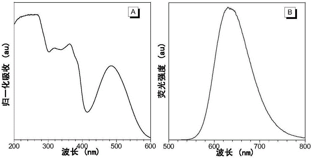
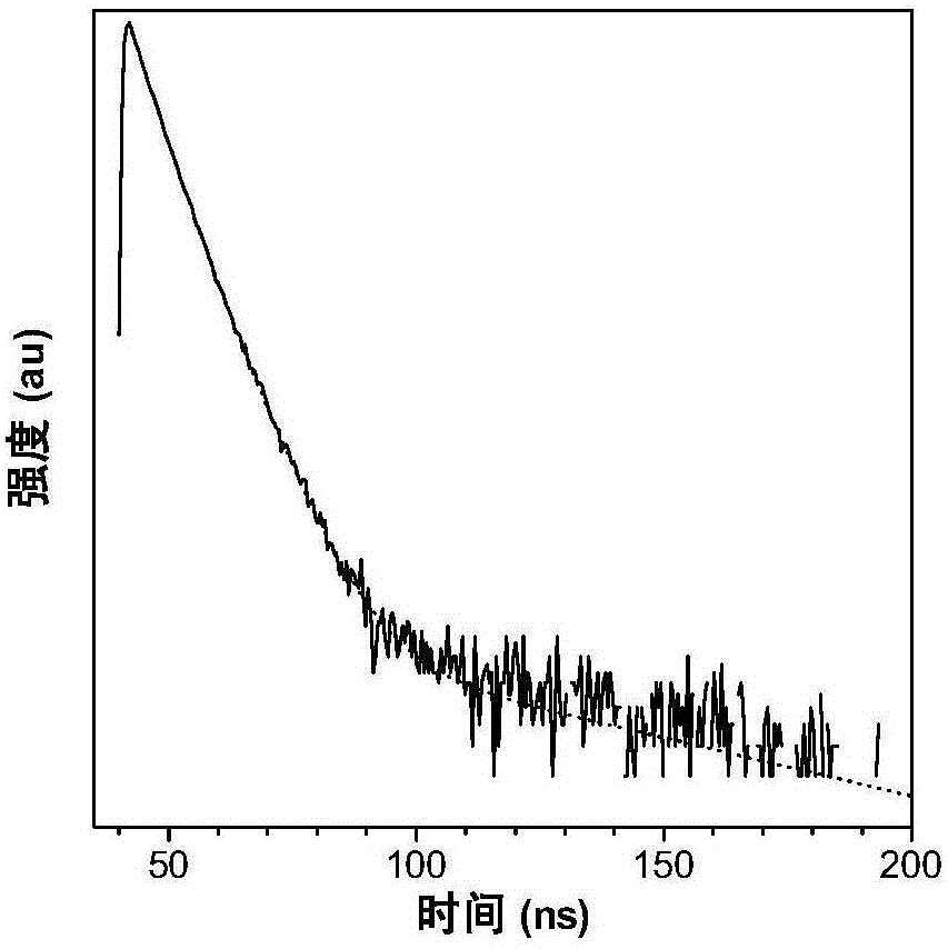
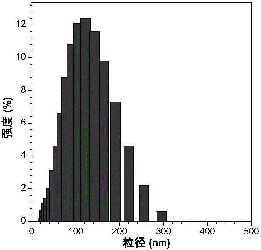
[0055] The nanoparticle t-BuPITBT-TPE-C225 has a maximum absorption at 475nm and a maximum emission at 625nm ( figure 1 ). The quantum yield is 35.1%, and the fluorescence lifetime is 4.63ns ( figure 2 ). The hydrated particle size of t-BuPITBT-TPE nanoparticles is 97nm, and the degree of dispersion (PDI) is 0.19 ( image 3 , measured by dynamic light scattering); the hydrated particle size of the t-BuPITBT-TPE-C225 nanoparticles modified with cetuximab is 116nm, and the degree of dispersion (PDI) is 0.33 ( Figure 4 ). It was further confirmed by TEM that the prepared fluorescent nanoparticles had good dispersion in aqueous solution ( Figure 5 and Figure 6 , Figure 5 was measured by dynamic light scattering).


PUM

PropertyMeasurementUnit
particle sizeaaaaaaaaaa
dispersityaaaaaaaaaa

Abstract

The invention discloses antibody-modified fluorescent nanoparticles and application thereof in targeted imaging of cancer cells. The fluorescent nanoparticles are prepared via the steps of dissolving an amphiphilic polymer and a fluorescent compound shown by formula I in a water-soluble organic solvent, and adding to water under ultrasonic action; blowing nitrogen to liquid surface to remove the organic solvent by volatilizing so as to form nanoparticles to which the fluorescent compound is supported; adding an antibody, 1-(3-dimethylaminopropyl)-3-ethylcarbodiimide hydrochloride and N-hydroxysulfosuccinimide into nanoparticle aqueous solution, and reacting to form amido bonds so as to prepare the fluorescent nanoparticles capable of targeted imaging. The traditional fluorescence imaging reagent has the defects such as quenching induced by aggregation, poor light stability and high cytotoxicity, while the fluorescent nanoparticles described herein have the advantages such as better detection results, high luminous efficiency, specific recognizing capacity, low cytotoxicity, good biocompatibility, and high light stability.

Description

technical field [0001] The invention belongs to the field of medical materials, in particular to an antibody-modified fluorescent nanoparticle and its application in targeted imaging of cancer cells. Background technique [0002] In cancer treatment, traditional chemotherapy usually has great side effects, which is due to the difficulty in distinguishing different cancer types at the molecular level by traditional cancer diagnostic methods, resulting in inefficient chemotherapy for patients with different cancer cell types method. [0003] In order to improve the treatment efficiency and reduce the side effects of the treatment process, it is necessary to diagnose cancer cells at the molecular level. Different cancer cells can be effectively distinguished by recognizing specific antigens on the surface of cancer cells. Monoclonal antibodies can specifically recognize and bind to antigens. For example, cetuximab can effectively bind to a variety of cancer cells that highly ...

Claims


Application Information

Patent Timeline
Patent Type & Authority Applications(China)
IPC IPC(8): C09K11/06G01N21/64
CPCC09K11/06C09K2211/1007C09K2211/1044C09K2211/1051C09K2211/1092G01N21/6486
Inventor 唐本忠高蒙赵祖金秦安军林耿伟
Owner SOUTH CHINA UNIV OF TECH
Why Eureka
Try Eureka