Enzyme linked immunosorbent assay kit for quantitatively detecting CD79 alpha

An enzyme-linked immunosorbent reagent, CD79 technology, applied in the biological field, can solve the problems of time-consuming and labor-intensive collection of samples, long time required, and inability to perform quantitative analysis.

Active Publication Date: 2016-11-23
广州瀚普创展医学检验实验室有限公司
View PDF3 Cites 4 Cited by
  • Summary
  • Abstract
  • Description
  • Claims
  • Application Information

AI Technical Summary

Problems solved by technology

[0003] The clinical method used to detect CD79α is mainly immunohistochemistry, but the experimental process of this method is more complicated, the time required is long, the sensitivity is low, quantitative analysis cannot be performed, and the reagents used are more harmfu

Method used


Image

  • Enzyme linked immunosorbent assay kit for quantitatively detecting CD79 alpha
  • Enzyme linked immunosorbent assay kit for quantitatively detecting CD79 alpha
  • Enzyme linked immunosorbent assay kit for quantitatively detecting CD79 alpha

Examples


Embodiment 1

[0025] Example 1: Preparation of polyclonal antibody

[0026] 1. Sources of gene and protein sequences

[0027] The human CD79α gene sequence can be found in XM_002829281 from NCBI.

[0028] Based on the gene sequence of CD79α, primers located upstream and downstream of the reading frame were synthesized.

[0029] Upstream primer: caccatggctgggg gtccaggagt cctcca

[0030] Downstream primer: ttctcgagtcacggcttc tccagctgga c

[0031] The full length of the human CD79α gene is 681bp, encoding 226 amino acids, of which amino acids 1 to 32 at the N-terminal are signal peptides. Protein sequence source: BAD97091 from NCBI.

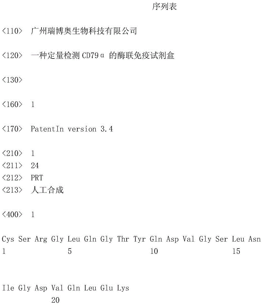
[0032] The polypeptide sequence used to prepare the monoclonal antibody is CSRGLQGTYQDVGSLNIGDVQLEK (SEQ ID NO.1).

[0033] 2. Construction of expression plasmids and acquisition of high-expression engineered strains

[0034] Expression vector construction method: obtain the target human CD79α gene by PCR amplification, use NdeI+XhoI double-cutting enzyme P...

Embodiment 2

[0042] Example 2. Preparation of CD79α monoclonal antibody

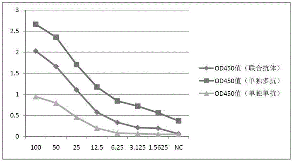
[0043] The monoclonal antibody was prepared by conventional methods: the peptide sequence CSRGLQGTYQDVGSLNIGDVQLEK (SEQ ID NO.1) used to prepare the monoclonal antibody was coupled with KLH and then immunized with SPF Balb / c mice, and the resulting mouse spleen was fused with myeloma cells SP2 / 0 and then screened Three hybridoma cell lines were obtained, and the antibodies secreted by the hybridoma cells were further purified with ProteinG / A affinity column to obtain anti-CD79α monoclonal antibody. One of the three hybridoma cell lines was selected by indirect ELISA and combined with the polyclonal antibody as the combined capture antibody.

Embodiment 3

[0044] Example 3: ELISA Kit for Detecting CD79α

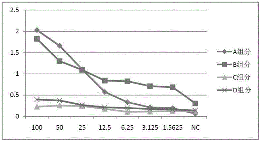
[0045] Set up the enzyme-linked immunosorbent assay kit for detecting CD79α, so that it contains the following components:

[0046] 1. Enzyme plate coated with anti-CD79α polyclonal antibody and anti-CD79α monoclonal antibody and used as capture antibody;

[0047] 2. pH value is 7.2, containing 5% skimmed milk powder, 0.01% Tween 20, trehalose, 0.1mol / L phosphate buffer as blocking solution;

[0048] 3. The pH value is 7.2, containing 0.5% Tween 20, 0.1mol / L washing solution of phosphate buffer;

[0049] 4. CD79α standard solution;

[0050] 5. The substrate chromogenic solution is composed of chromogenic solution A and chromogenic solution B, chromogenic solution A is hydrogen peroxide, and chromogenic solution B is o-phenylenediamine or tetramethylbenzidine;

[0051] 6. The stop solution is 2M sulfuric acid solution;

[0052] 7. Biotinylated anti-CD79α monoclonal antibody (Catalog No. 130-10122) as detection antibody

[0...


PUM

PropertyMeasurementUnit
Linearaaaaaaaaaa

Abstract

The invention relates to an enzyme linked immunosorbent assay kit for quantitatively detecting CD79 alpha. The enzyme linked immunosorbent assay kit for quantitatively detecting CD79 alpha comprises an enzyme-labeled panel coated with a joint capture antibody, confining liquid, washing liquid, a CD79 alpha standard product solution, substrate developing liquid, stopping liquid, an anti-CD79 alpha monoclonal antibody detection antibody and streptavidin labeled by pepper peroxidase. The joint capture antibody comprises an anti-CD79 alpha polyclonal antibody and an anti-CD79 alpha monoclonal antibody obtained by a fusion protein immunological experiment animal. The enzyme linked immunosorbent assay kit is easy to operate, short in time, high in sensitivity and suitable for rapid detection of a large number of samples.

Description

technical field [0001] The invention belongs to the field of biotechnology, and relates to an enzyme-linked immunoassay kit for detecting CD79α and a preparation method thereof. Background technique [0002] CD79α is a member of the immunoglobulin superfamily (IgSF). It is positively expressed in B-cell lymphoma, most acute B-lymphoblastic leukemia and myeloma. As an auxiliary detection factor of CD20, it is widely used in the identification of B-cells and their source tumors. The positive sites of CD79α expression were in the membrane and cytoplasm. CD79α is a broad-spectrum marker of B cells, from pre-B initiation to mature plasma cells. CD79α will have a partial immune response crossover with CD79β during the regulation of B cell terminal differentiation. CD79α can be used to differentiate B-cell lymphoma CD79α(+), pre-B-cell acute lymphoblastic leukemia (ALL) CD79α(+) and acute T-cell lymphoblastic leukemia CD79α(-). [0003] The clinical method used to detect CD79α ...

Claims


Application Information

Patent Timeline
IPC IPC(8): G01N33/68
Inventor 罗树红黄若磐
Owner 广州瀚普创展医学检验实验室有限公司
Why Eureka
Try Eureka