Leucodermia virus rapid detecting kit, and its preparing and using method

A technology for detecting kits and viruses, applied in the interdisciplinary field of immunology and virology, to achieve the effects of simplified detection steps, shortened time, and improved sensitivity

Active Publication Date: 2005-07-20
OCEAN UNIV OF CHINA
View PDF0 Cites 4 Cited by
  • Summary
  • Abstract
  • Description
  • Claims
  • Application Information

AI Technical Summary

Problems solved by technology

The application of this method in the detection of pathogens of aquaculture diseases is still blank

Method used


Image

  • Leucodermia virus rapid detecting kit, and its preparing and using method
  • Leucodermia virus rapid detecting kit, and its preparing and using method
  • Leucodermia virus rapid detecting kit, and its preparing and using method

Examples


Embodiment 1

[0036]The colloidal gold labeling method of described two kinds of monoclonal antibodies E and D is:

[0037] (1) Preparation of colloidal gold: preparation of 18-20nm colloidal gold particles, mixing 100ml of 0.01% chloroauric acid solution with 2.5ml of 0.1% sodium citrate, heating to 100°C to make a colloidal gold solution, the pH value of the solution: Adjust to 8.2-8.3 with 0.2% potassium carbonate, set aside;

[0038] (2) Preparation of gold-labeled antibody: Add appropriate amount of monoclonal antibody E and monoclonal antibody D to the colloidal gold solution of the above (1), stir slowly for 10 minutes, add 1% bovine serum albumin, and overnight at 4°C; It was centrifuged at 18000g for 110min at 4°C; the centrifuged precipitate was washed with 0.01M phosphate buffered saline (PBS: KCI 0.2g, NaCl 8.0g, KH 2 PO 4 0.2g, Na 2 HPO 4 12H 2 O 2.9g, distilled water 1000ml, pH 7.4) to suspend, the dosage is one-tenth of the original volume. Prepare gold-labeled monocl...

Embodiment 2

[0045] The appropriate amount of monoclonal antibody is: when the concentration of monoclonal antibody is 0.5g / L, the optimal amount of monoclonal antibody labeled with colloidal gold per milliliter is: 15-20 μl. Add 1 μl of antibody to 1ml of colloidal gold, mix well, add 100 μl of 10% sodium chloride, observe the color change, if it turns blue, it means that the antibody is insufficient, and then continue to increase the amount of antibody until the color of the colloidal gold does not change. On this basis, increase the amount of this antibody by 50-100%, which is the appropriate amount of monoclonal antibody at this time. When the appropriate amount of monoclonal antibody is used for colloidal gold labeling, the obtained colloidal gold probe has no precipitation and no non-specific adsorption, which reaches the experimental standard.

Embodiment 3

[0047] The hypotonic solution A with an osmotic pressure of less than 360mOsm / Kg consists of: distilled water; or tap water; or phosphate buffer with pH 7.0-7.6; or Tris-sodium chloride buffer with pH 7.0-7.6.

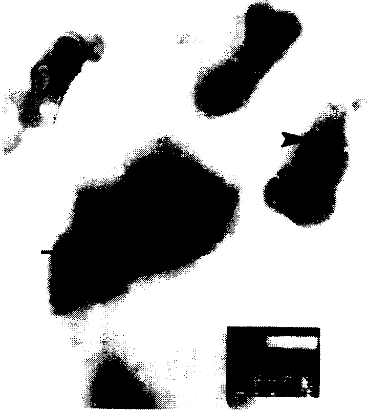
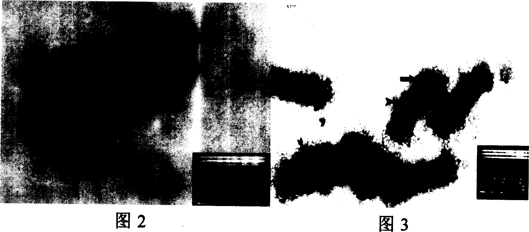
[0048] The hypotonic solution of the present invention has an osmotic pressure of less than 360mOsm / Kg, which is lower than the 389-410mOsm / Kg blood osmotic pressure of the freshwater animal-crayfish, and 600mOsm / Kg lower than the 600mOsm / Kg blood osmotic pressure of the seawater animal-Penaeus sinensis. The envelope of white spot disease virus isolated from shrimp blood is complete. Therefore, shrimp blood is the isotonic fluid of white spot disease virus. Because the envelope of white spot disease virus is similar to biofilm, in low osmotic fluid, at osmotic pressure Under the action, the water outside the capsule enters the capsule, causing the capsule to burst. The capsule of white spot virus is ruptured in the liquid with osmotic pressure less than 360mOsm / Kg, see...


PUM


Abstract

The invention is a melanodermia virus quickly-detecting reagent box, including: a detecting device; tween-phosphate buffer liquid containing ox serum albumin; tween-phosphate buffer liquid; low osmotic liquid; gold labeled anti-melanodermia virus monoclonal antibody probe, prepared of two colloidal gold labeled monoantibodies E and D in proportion; the E and D are anti-melanodermia virus bursa membrane antibody E and anti-melanodermia virus nucleus coat antibody D secreted by two hybrid tumor cells, respectively; the conservation numbers of the two hybrid tumor cells are CCTCC-C200421 and CCTCC-C200422, respectively. As the E and D are used as detecting antibodies, they can obviously improve detecting sensitivity. The detecting reaction only takes 3 minutes without hatching and any instrument and equipment, and their detecting sensitivity is the same as that of spot immune printing method. The reagent box has the characters: simple and convenient, quick, accurate, etc.

Description

technical field [0001] The invention relates to the improvement of the detection technology of breeding disease pathogens, specifically a white spot syndrome virus (white spot syndrome virus, WSSV) rapid detection kit and its preparation and use method, which belong to the interdisciplinary technical field of immunology and virology. Background technique [0002] White spot virus disease is one of the serious diseases in prawn farming, which leads to high mortality of prawns and huge losses to the prawn farming industry. Given that there is no effective treatment for leukoplakia, rapid and accurate virus isolation and detection technology has become one of the focuses of research by scholars from all over the world. At present, the diagnosis of leukoplakia virus disease mainly depends on the detection of leukoplakia virus in the laboratory. The current laboratory detection methods include: PCR method; DNA probe in situ hybridization method; transmission electron microscope o...

Claims


Application Information

Patent Timeline
Patent Type & Authority Applications(China)
IPC IPC(8): G01N33/52G01N33/532G01N33/544G01N33/569
Inventor 战文斌王晓洁
Owner OCEAN UNIV OF CHINA
Why Eureka
Try Eureka