Preparation method and applications of decellularized collagen gel microsphere scaffold

A collagen gel microsphere, collagen gel technology, applied in medical science, tissue regeneration, prosthesis, etc., can solve the problems of inability to retain the biological properties of decellularized scaffolds, matrix and factor damage, and achieve favorable adhesion and Effect of tissue healing, low rejection, avoidance of fragmentation process

Inactive Publication Date: 2017-07-11
SICHUAN UNIV
View PDF9 Cites 7 Cited by
  • Summary
  • Abstract
  • Description
  • Claims
  • Application Information

AI Technical Summary

Problems solved by technology

Excessive decellularization will lead to the destruction of matrix and factors, and the biological performance of decellularized scaffolds cannot be preserved

Method used


Image

  • Preparation method and applications of decellularized collagen gel microsphere scaffold
  • Preparation method and applications of decellularized collagen gel microsphere scaffold
  • Preparation method and applications of decellularized collagen gel microsphere scaffold

Examples


Embodiment 1

[0027] Prepare collagen gel microspheres loaded with bone marrow mesenchymal stem cells as follows

[0028] 1. Take a collagen solution with a concentration of 15mg / mL, adjust the pH to 7.0 with a small amount of 1mol / L sodium hydroxide solution in an ice-water bath, and dilute the collagen solution to 8mg / mL;

[0029] 2. Count the bone marrow mesenchymal stem cells prepared in advance and prepare a cell suspension with α-MEM medium, add it to the collagen solution and disperse evenly, the final concentration of collagen is 6.5mg / mL, and the cell density is 5× 10 6 a / mL;

[0030] 3. Select 70mL of methyl silicone oil with a viscosity of 100mPa·s, add 0.05% (v / v) Span-80, and stir with a magnetic stirrer at a speed of 500rpm;

[0031] 4. Add the cell-collagen mixture dropwise to the oil phase in an ice-water bath, and continue stirring for 30 minutes; then raise the temperature of the water-oil phase mixture system to 37 degrees, and continue stirring for 20 minutes; after th...

Embodiment 2

[0034] Prepare chondrocyte-loaded collagen gel microspheres as follows

[0035] 1. Take a collagen solution with a concentration of 15mg / mL, adjust the pH to 7.0 with a small amount of 1mol / L sodium hydroxide solution in an ice-water bath, and dilute the collagen solution to 8mg / mL;

[0036] 2. Count the chondrocytes prepared in advance and prepare a cell suspension with α-MEM medium, add it to the collagen solution and disperse evenly. The final concentration of collagen is 5mg / mL, and the cell density is 1×10 7 a / mL;

[0037] 3. Select 70mL of methyl silicone oil with a viscosity of 100mPa·s, add 0.1% (v / v) Span-80, and stir with a magnetic stirrer at a speed of 600rpm;

[0038] 4. Add the cell-collagen mixture dropwise to the oil phase in an ice-water bath, and continue stirring for 30 minutes; then raise the temperature of the water-oil phase mixture system to 37 degrees, and continue stirring for 20 minutes; after the stirring is completed, put Centrifuge the water phas...

Embodiment 3

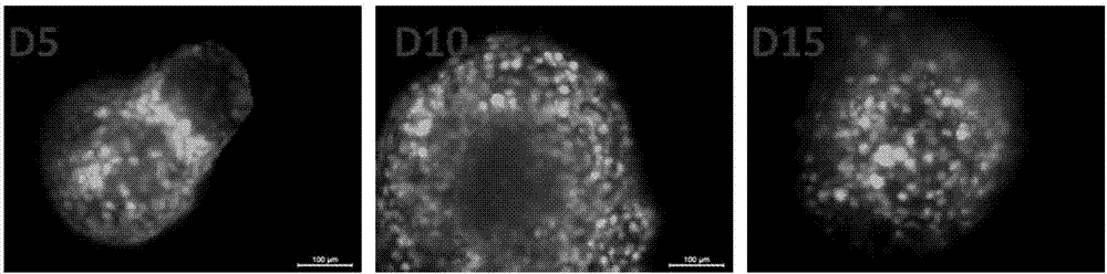
[0041] In vitro culture experiment of bone marrow mesenchymal stem cells-collagen gel microspheres

[0042]Bone marrow mesenchymal stem cells-collagen gel microspheres prepared in Example 1 were cultured in chondrogenic medium for 5 days, 10 days and 15 days, and the samples were stained with fluorescein diacetate (FDA) to observe the state of the cells. The result is as figure 2 shown.

[0043] from figure 2 It can be seen that the bone marrow mesenchymal stem cells-collagen gel microspheres prepared in Example 1 maintained a spherical shape from day 5, indicating that the stem cells had differentiated into chondrocytes, and the cell density gradually decreased. The size of the microspheres shrunk a little on the first day, because the cells began to adhere and did not differentiate into chondrocytes at this time, and the size was basically stable after the fifth day.


PUM

PropertyMeasurementUnit
viscosityaaaaaaaaaa
diameteraaaaaaaaaa

Abstract

The invention provides a decellularized collagen gel microsphere scaffold preparation method, and belongs to the technical field of decellularized scaffold tissue engineering. The decellularized collagen gel microsphere scaffold preparation method comprises preparation of a cell-collagen mixing liquid, preparation of cell-collagen gel microspheres, and preparation of a decellularized collagen gel microsphere scaffold. According to the present invention, by directly preparing the cell coating collagen gel microspheres, the micrometer-scale tissue block is obtained through in vitro culture, such that the decellularization operation can be directly performed while a large amount of the micrometer-scale tissue can be obtained, the subsequent breaking process is avoided, and the matrix and the micro-structure in the tissue are well retained; the decellularized collagen gel microsphere scaffold does not have DNA residue so as to minimize the rejection reaction; and the size of the decellularized collagen gel microsphere scaffold is micrometer-scale, and after the decellularized collagen gel microsphere scaffold is used in cartilage and subchondral bone defect repair, the cell adhesion and the tissue healing are easily achieved, the uniformly distributed tissue is formed at the late stage, the hollowing can be avoided, and the probability of surgical trauma and late stage infections can be reduced.

Description

technical field [0001] The invention belongs to the technical field of decellularized scaffold tissue engineering, in particular to a preparation method and application of decellularized collagen gel microsphere scaffold. Background technique [0002] Acellular scaffolds are common tissue engineering scaffolds. The sources of decellularized scaffolds mainly include natural tissue and in vitro tissue engineering. But there are some problems. Due to source issues, human-derived natural tissues are relatively rare. Most of them use heterogeneous sources, and products such as pig eye cornea and bovine pericardium are very common. However, tissues of xenogeneic origin are more prone to rejection. In vitro tissue engineering can also obtain tissues that meet certain functions, such as fat, cartilage, and bone, which are easier to obtain and mass-produce than natural sources. Decellularization is the most critical step in the preparation of decellularized scaffolds. The remova...

Claims


Application Information

Patent Timeline
Patent Type & Authority Applications(China)
IPC IPC(8): A61L27/36A61L27/24A61L27/50A61L27/52
CPCA61L27/24A61L27/3654A61L27/3687A61L27/3691A61L27/50A61L27/52A61L2300/412A61L2430/06A61L2430/40
Inventor 樊渝江刘钧林海王启光孙勇
Owner SICHUAN UNIV
Why Eureka
Try Eureka