Circulating tumor cell separation micro-fluidic chip device and application method thereof

A microfluidic chip and tumor cell technology, which is applied in the field of devices for automatic sorting of circulating tumor cells, can solve the problems of missed detection of small particle size tumor cells, complex chip preparation, and many operation procedures, and achieves a high degree of automation and a chip. Simple structure, overcoming the effect of low integration

Pending Publication Date: 2017-08-22
上海纳奥生物科技有限公司
View PDF12 Cites 23 Cited by
  • Summary
  • Abstract
  • Description
  • Claims
  • Application Information

AI Technical Summary

Problems solved by technology

The overall preparation of the chip is complicated, the operation process is many and the cost is high
[0006] Patent 201410345305.6 discloses a double-helix microfluidic chip, which includes red blood cell outlets, white blood cell outlets, fluid outlets, and cell filter membranes. Fundamentally, it still relies on 8-micron pore size filter membranes to achieve filtration and separation of tumor cells. Such methods still suffer from Missed small-sized tumor cells

Method used


Image

  • Circulating tumor cell separation micro-fluidic chip device and application method thereof
  • Circulating tumor cell separation micro-fluidic chip device and application method thereof
  • Circulating tumor cell separation micro-fluidic chip device and application method thereof

Examples


Embodiment

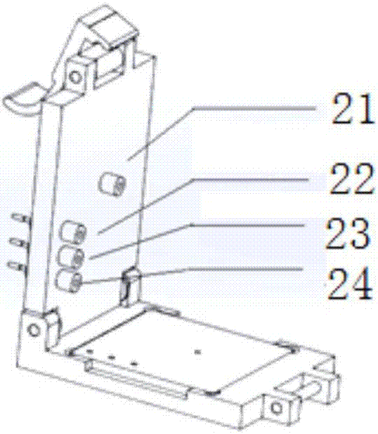
[0033] A microfluidic chip device for separating circulating tumor cells, the device includes a microfluidic chip 1 and a chip holder 2 .

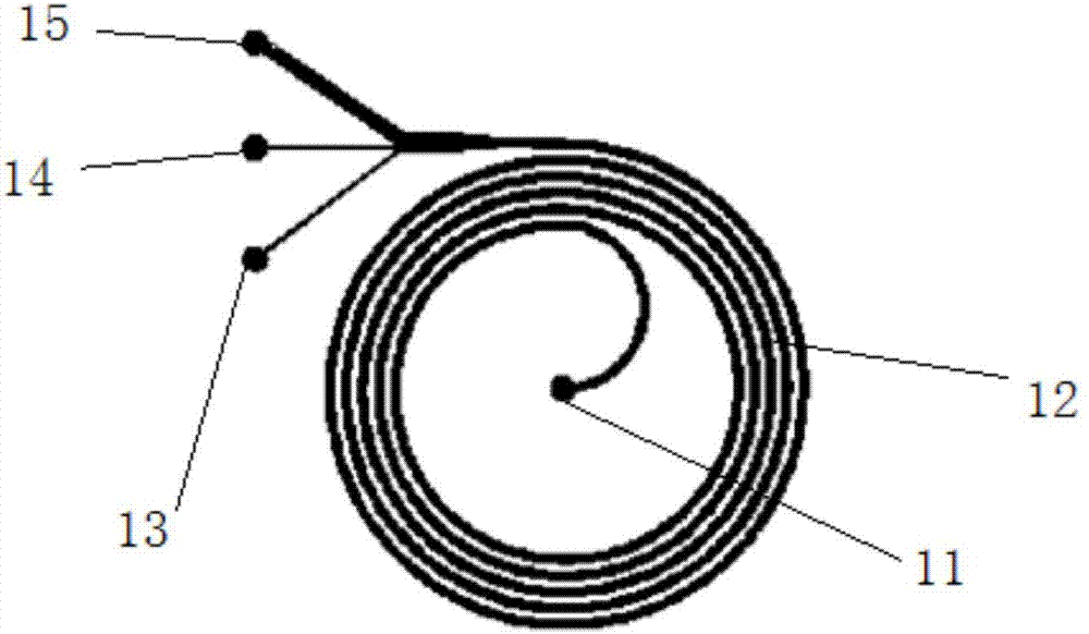
[0034] Such as figure 1Said, the microfluidic chip 1 includes a chip inlet 11, a single helix chip 12, a rare cell collection pipeline and a white blood cell outlet 15, wherein the rare cell collection pipeline includes two, respectively connected to the inner outlet 13 and the outlet 14 The single helical chip 12 is composed of a single helical channel, the single helical channel enters the helical microfluidic channel from the chip inlet 11 in the center of the circular helical channel through the semicircular initial channel, and the end contains 3 outlets: the inner outlet 13 and outlet 14 for collecting rare cells of different particle sizes, and the outer white blood cell outlet 15 for collecting white blood cells; wherein, the width of the inner outlet 13 is 110±50 μm, and the width of the outlet 14 is 110 ±50 μm, the width of the ...

Embodiment 2

[0046] The microfluidic device was used to separate circulating tumor cells in pleural effusion, the device was the same as in Example 1, and the method was as follows:

[0047] (1) The pleural effusion sample was gently turned upside down back and forth to make it evenly mixed; the pleural effusion was diluted 100 times with PBS, and sucked with a 30ml syringe; The interface of the control chip device is connected, and the flow rate of the pleural effusion pump is 130ml / h.

[0048] (2) The enrichment separation liquid flows out from the enrichment separation outlet and is put into a 5ml sterile centrifuge tube.

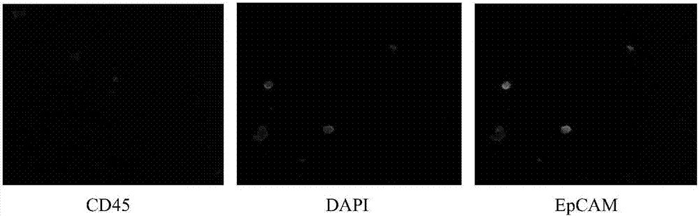
[0049] (3) Spin the circulating tumor cells separated by microfluidics to make sample slides. Fix with 4% paraformaldehyde, perform Giemsa staining, observe and take pictures under a microscope, such as Figure 7 As shown, the isolated tumor cells showed typical cell morphology with large nuclei, high nuclear-to-cytoplasmic ratio, and large particle size.

Embodiment 3

[0051] Both the inner outlet 13 and the outlet 14 are surface-coated with positively charged polymer acrylamide. The biological sample is injected into the chip device at a constant rate through a syringe pump or a pressure pump device, and the flow rate is 50ml / h. The volume ratio of the PBS buffer solution to the blood sample to be tested is 10:1. All the other are with embodiment 1.


PUM

PropertyMeasurementUnit
widthaaaaaaaaaa
widthaaaaaaaaaa
widthaaaaaaaaaa

Abstract

The invention relates to a circulating tumor cell separation micro-fluidic chip device and an application method thereof. The device comprises a micro-fluidic chip (1) and a chip fixture (2). The micro-fluidic chip (1) comprises a chip inlet (11), a single-screw chip (12), rare cell collection tubes (13, 14) and a leucocyte outlet (15). A single-screw channel starts from the chip inlet (11) at the center of a circular spiral channel, passes through a semicircular starting channel and enters a spiral micro-fluidic channel. The tail end contains three outlets: rare cells with different particle sizes are respectively collected; and the chip fixture (2) is automatically in butt joint with inlet and outlet of the micro-fluidic chip (1). In comparison with the prior art, the micro-fluidic chip device of the invention has the following beneficial effects: a treated sample can be automatically subjected to automated high-throughput tumor cell separation and tumor cells in a liquid sample such as peripheral blood/pleural effusion/cerebrospinal fluid, etc. of a cancer patient can be enriched; and cancer patient diagnosis and customized treatment specificity and accuracy are remarkably enhanced.

Description

technical field [0001] The invention relates to the field of cell sorting, in particular to a device and method for automatically sorting circulating tumor cells from biological liquid samples on a microfluidic chip. Background technique [0002] In fields such as biomedicine, it is often necessary to sort out specific target cells from more complex samples. For example, detecting, sorting and counting the number of circulating tumor cells (CTC, circulating tumor cells) in the blood of cancer patients is becoming more and more important in the diagnosis and treatment of cancer. At present, CTC detection techniques are mixed, generally using and not using markers. Marker-based capture usually includes positive enrichment and negative enrichment based on antibody-coated magnetic beads. This category belongs to the first generation of CTC capture technology, represented by Johnson & Johnson's Cell search system. However, its technical defect is that antibody capture is limite...

Claims


Application Information

Patent Timeline
Patent Type & Authority Applications(China)
IPC IPC(8): G01N15/10
CPCG01N15/1056G01N2015/1081
Inventor 亓立峰
Owner 上海纳奥生物科技有限公司
Why Eureka
Try Eureka