

Analysis method for detecting drug distribution and application thereof
An analysis method and drug technology, applied in the direction of analyzing materials, material analysis by electromagnetic means, measuring devices, etc., can solve the problems of difficult to truly reflect the distribution of drugs, unable to detect the distribution of drugs, etc., and achieve the effect of accurate drug distribution information.
- Summary
- Abstract
- Description
- Claims
- Application Information
AI Technical Summary
Problems solved by technology
Method used
Image

Examples
Embodiment 1
[0050] The present embodiment provides an analytical method for detecting drug distribution, which includes:
[0051] Step a. Construct an in vitro myocardial tissue model: take the sterilized decellularized mouse myocardial tissue and place it in a 24-well plate whose inner wall is pre-coated with fibronectin; put the medium containing cardiomyocytes at a concentration of 50,000 cells / mL Inject the suspension into a 24-well plate; place the 24-well plate in an incubator for 4 hours, continue to add medium, and co-culture cardiomyocytes with decellularized mouse myocardial tissue for 7 days to obtain an in vitro myocardial tissue model.
[0052] Step b. Administration: the isotope 13 The C-labeled drug to be tested was added to the culture medium of the myocardial tissue model in vitro, and cultured for 48 hours under the same conditions.
[0053] Step c. Preparation and detection of samples to be tested: use 2.5% glutaraldehyde solution to chemically fix the in vitro myocard...
Embodiment 2
[0055] The present embodiment provides an analytical method for detecting drug distribution, which includes:
[0056] Step a. Constructing an in vitro stem cell co-culture model: take the sterilized decellularized porcine small intestinal mucosa tissue and place it in a 24-well plate whose inner wall is pre-coated with fibronectin; Inject the culture medium suspension into the 24-well plate; place the 24-well plate in the incubator for 4 hours, continue to add the medium, co-culture the mesenchymal stem cells with the decellularized porcine small intestinal mucosa tissue for 7 days, and obtain the in vitro Stem cell co-culture model.
[0057] Step b. Administration: the isotope 15 The N-labeled drug to be tested was added to the culture medium of the in vitro stem cell co-culture model, and cultured for 72 hours under the same conditions.
[0058] Step c. Preparation of the sample to be tested: After the in vitro stem cell co-culture model obtained in step b is subjected to ...
experiment example 1
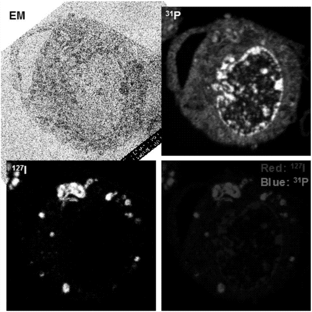
[0061] Distribution detection of amiodarone in macrophages:
[0062] 1. Experimental method
[0063] 1. Construction of in vitro macrophage co-culture model: Decellularized natural soft tissue and macrophages (NR8383) were co-cultured in a culture dish.
[0064] 2. Administration: Amiodarone solution with a concentration of 1.56 μg / mL was added to the macrophage culture medium, and in 5% CO 2 and 37°C for 72 hours.
[0065] 3. Sample processing: use the chemical fixation method to process the co-culture model of macrophages after administration, that is, a solution prepared with 0.5% tannic acid and 2.5% glutaraldehyde (adjusted with 0.08m sodium cacodylate) pH to 7) to treat the macrophage co-culture model, followed by embedding with epoxy resin. After the resin was cured, cut into 500nm thin slices with a microscope microtome and dried on the surface of the silicon wafer.
[0066] 4. Electron microscope imaging: use a scanning electron microscope to detect the dried flak...
PUM
 Abstract
Description
Claims
Application Information