Method for detecting haptoglobin (Hp) classified type

A technology of haptoglobin and polymerase, which is applied in the field of biomedicine and can solve problems such as low accuracy rate and complex typing methods for detecting haptoglobin

Active Publication Date: 2017-11-28
深圳中科唯新生物技术有限公司 +2
0 Cites 0 Cited by

AI-Extracted Technical Summary

Problems solved by technology

[0009] The present invention provides a method for detecting haptoglobin typing, to at least solve ...

Method used

A kind of method that the embodiment of the present invention 1-3 establishes to analyze Hp from the genetic angle, said method carries out gene amplification by designing specif...

Abstract

The invention provides a method for detecting the haptoglobin (Hp) type. According to the method, by designing specific primers, a to-be-detected sample containing Hp genes is subjected to gene amplification, and thus amplified products are obtained, and by conducting electrophoresis detection on the amplified products, the gene type of Hp of the to-be-detected sample can be determined. The method that type classifying is conducted on the Hp from the perspective of genes is built, and operation of the method is easy, convenient, fast and accurate so that the method can be applied to clinical practice.

Application Domain

Microbiological testing/measurementDNA/RNA fragmentation

Technology Topic

BiologyGenotype +4

Image

  • Method for detecting haptoglobin (Hp) classified type
  • Method for detecting haptoglobin (Hp) classified type
  • Method for detecting haptoglobin (Hp) classified type

Examples

  • Experimental program(4)

Example Embodiment

[0051] Example 1:
[0052] Step 1: Saliva sample collection and processing
[0053] Rinse mouth 30 minutes before sampling to remove food residues and residual microorganisms, do not eat, drink, brush teeth, smoke or chew gum within 30 minutes after rinsing; relax cheeks and rub gently for 15-30 seconds to generate saliva, collect 2ml of saliva into a sterile collector.
[0054] Step 2: DNA (deoxyribonucleic acid) extraction from saliva samples
[0055] (1) Take 500ul (microliter) saliva sample, add 1ml (ml) buffer (100mmol/L (mmol/L) Tris-HCl (tris-hydrochloric acid buffer), pH8.0, 0.5 % SDS (sodium dodecyl sulfonate), 10mmol/LEDTA (ethylenediaminetetraacetic acid)) and 6ul 20mg/ul (mg/microliter) proteinase K, shake and mix on a vortexer;
[0056] (2) the saliva treated in step (1) is treated in a water bath at 55°C for 20min;
[0057] (3) Add 600ul of phenol:chloroform:isoamyl alcohol (25:24:1), invert and mix, centrifuge at 10000rpm (revolution/min) for 5min, and take the supernatant;
[0058] (4) adding 600ul of chloroform:isoamyl alcohol (24:1) to the supernatant of step (3), inverting and mixing, and centrifuging at 10,000 rpm for 5 min, and taking the supernatant;
[0059] (5) adding equal volume of isopropanol to the supernatant liquid of step (4), gently mixing and centrifuging at 14000rpm for 10min, discarding the supernatant;
[0060] (6) Wash the above-mentioned precipitate once with 500ul of 70% ethanol, centrifuge at 14000rpm for 10min, and discard the supernatant;
[0061] (7) Air-dry DNA at room temperature and dissolve in 20ul of sterile water.
[0062] Step 3: Gene PCR (polymerase chain reaction) amplification and product identification
[0063] (1) PCR amplification:
[0064] Use 2X Mix buffer containing Tris-HCl (final concentration: 10mmol/L), pH 8.3, potassium chloride (final concentration: 50mmol/L), magnesium chloride (final concentration: 1.5mmol/L), Taq enzyme (final concentration: 2U), a mixed solution of dNTPs (final concentration: 200 umol/L), and the DNA extracted from the saliva sample was used as a template for PCR amplification.
[0065] The amplification primers are as follows:
[0066] F: 5'-ATAATACAGTTCGCGAGCTTCT-3';
[0067] R: 5'-GGGCAAGATACTCAACCTGTC-3'.
[0068] The PCR reaction program is as follows:
[0069] 95℃ for 4min; 95℃ for 40s, 60℃ for 30s, 72℃ for 80s, 30 cycles; 72℃ for 3min; 4℃∝.
[0070] That is, pre-denaturation at a temperature of 95°C for 4 minutes; denaturation at a temperature of 95°C for 40 seconds, annealing at a temperature of 60°C for 30 seconds, and extension at a temperature of 72°C for 80 seconds, wherein the denaturation step, the The annealing step and the extension step were 30 cycles; then a final extension at a temperature of 72°C for 3 minutes; the final hold temperature was 4°C.
[0071] (2) Detection by agarose gel electrophoresis
[0072] see the results figure 1 , figure 1 An image of agarose gel after electrophoresis provided in Example 1 of the present invention;
[0073] Lanes 2 to 4 are positive control samples with known Hp genotypes, and lane 5 is the sample to be tested; lane 2 is Hp2-2 subtype, lane 3 is Hp2-1 subtype, and lane 4 is Hp1-1 subtype type. Lane 1 is DNA marker (DNA marker) (the bands are 100bp, 250bp, 500bp, 750bp, 1kb, 1.5kb, 2kb, 3kb, 5kb from bottom to top).
[0074] Step 4: Determine the genotype of Hp
[0075] According to the electrophoresis results to determine the genotype of Hp, in theory, the Hp1-1 genotype has no band, the Hp2-1 genotype band is 698bp, and the Hp2-2 genotype band is 698bp. It can be seen that the Hp genotype of the sample to be tested in this example is the Hp2 genotype.
[0076] The method for detecting haptoglobin typing established by the primers provided in this example can better distinguish Hp1 subtype and Hp2 subtype. Since Hp1-1 has no band, there are two bands that should exist in Hp2-1. Hp2-1 and Hp2-2 cannot be identified from the electropherogram, but the method for detecting haptoglobin typing established by the primers provided in this example can better distinguish Hp1 subtype and Hp2 Subtype, and the operation is extremely simple, and the test results are accurate. For diabetic patients who only need to distinguish the Hp1 subtype genotype, it is determined that vitamin E cannot be supplemented, otherwise it will increase the risk of cardiovascular disease. Therefore, Primers that can only differentiate between Hp1 subtypes and Hp2 subtypes also have clinical value.

Example Embodiment

[0077] Example 2:
[0078] Step 1: Peripheral blood sample processing and DNA extraction
[0079] (1) Take 2ml of peripheral blood containing EDTA anticoagulant, put it in a 10ml sterilized centrifuge tube, add 6mL of 0.1mM EDTA, process for 15 minutes to disrupt red blood cells, centrifuge at 3000rpm for 10min, discard the supernatant, and collect the precipitate;
[0080] (2) Add 1 ml of 0.1 mM EDTA to the precipitate, mix well and transfer it to a 2 ml sterilized centrifuge tube, centrifuge at 5400 rpm for 5 min, and discard the supernatant;
[0081] (3) Add 1ml buffer (100mmol/LTris-HCl, pH8.0, 0.5%SDS, 10mmol/LEDTA) and 6ul 20mg/ul proteinase K to the precipitate, shake and mix on a vortexer, and then treat in a water bath at 55°C 20min;
[0082] (4) Add 600ul of phenol:chloroform:isoamyl alcohol (25:24:1), invert and mix, centrifuge at 10000rpm for 5min, and take the supernatant;
[0083] (5) adding 600ul of chloroform:isoamyl alcohol (24:1) to the supernatant of step (4), inverting and mixing, and centrifuging at 10,000 rpm for 5 min, and taking the supernatant;
[0084] (6) adding equal volume of isopropanol to the supernatant liquid of step (5), gently mixing and centrifuging at 14000rpm for 10min, discarding the supernatant;
[0085] (7) Wash the above-mentioned precipitate once with 500ul of 70% ethanol, centrifuge at 14000rpm for 10min, and discard the supernatant;
[0086] (8) Air-dry DNA at room temperature and dissolve in 30ul of sterile water.
[0087] Step 2: Gene PCR (polymerase chain reaction) amplification and product identification
[0088] (1) PCR amplification:
[0089] Use 10X Mix buffer containing Tris-HCl (final concentration: 8mmol/L), pH 8.0, potassium chloride (final concentration: 60mmol/L), magnesium chloride (final concentration: 1.0mmol/L), Taq enzyme (final concentration: 2.5U), a mixed solution of dNTPs (final concentration: 110 umol/L), and the DNA extracted from the saliva sample was used as a template for PCR amplification.
[0090] The amplification primers are as follows:
[0091] F: 5'-ATAATACAGTTCGCGAGCTTCT-3';
[0092] R: 5'-GGGCAAGATACTCAACCTGTC-3'.
[0093] The PCR reaction program is as follows:
[0094] 98℃ for 5min; 98℃ for 35s, 63℃ for 22s, 70℃ for 60s, 38 cycles; 70℃ for 8min; 3℃∝.
[0095] That is, pre-denaturation at a temperature of 98°C for 5 minutes; denaturation at a temperature of 98°C for 35 seconds, annealing at a temperature of 63°C for 22 seconds, and extension at a temperature of 70°C for 60 seconds, wherein the denaturation step, the The annealing step and the extension step were 38 cycles; then a final extension at a temperature of 70°C for 8 minutes; the final hold temperature was 3°C.
[0096] (2) Detection by agarose gel electrophoresis
[0097] see the results figure 2. figure 2 An image of agarose gel after electrophoresis provided in Example 2 of the present invention;
[0098] Among them, lane 1 is the sample to be tested, lanes 2 and 3 are control samples, lane 2 is Hp2-1 subtype, lane 3 is Hp1-1 subtype, and lane 4 is DNA Marker (the bands are 100bp from bottom to top). , 200bp, 300bp, 400bp, 500bp, 600bp, 700bp, 800bp, 900bp, 1kb, 1.5kb).
[0099] Step 3: Determine the genotype of Hp
[0100] According to the electrophoresis results to determine the genotype of Hp, in theory, the Hp1-1 subtype has no band, the Hp2-1 genotype band is 698bp, and the Hp2-2 genotype band is 698bp. It can be seen that the Hp genotype of the sample to be tested in this example is the Hp2 genotype.
[0101]The method for detecting haptoglobin typing established by the primers provided in this example can better distinguish Hp1 subtype and Hp2 subtype. Since Hp1-1 has no band, there are two bands that should exist in Hp2-1. Hp2-1 and Hp2-2 cannot be identified from the electropherogram, but the method for detecting haptoglobin typing established by the primers provided in this example can better distinguish Hp1 subtype and Hp2 Subtype, and the operation is extremely simple, and the test results are accurate. For diabetic patients who only need to distinguish the Hp1 subtype genotype, it is determined that vitamin E cannot be supplemented, otherwise it will increase the risk of cardiovascular disease. Therefore, Primers that can only differentiate between Hp1 subtypes and Hp2 subtypes also have clinical value.

Example Embodiment

[0102] Example 3:
[0103] Step 1: Same as step 1 in Example 2.
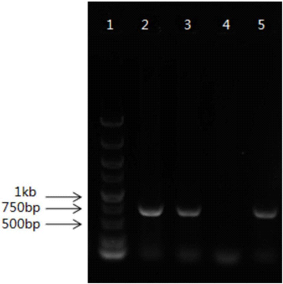
[0104] Step 2: Gene PCR (polymerase chain reaction) amplification and product identification
[0105] (1) PCR amplification:
[0106] Use 10X Mix buffer containing Tris-HCl (final concentration: 12mmol/L), pH 8.6, potassium chloride (final concentration: 40mmol/L), magnesium chloride (final concentration: 1.8mmol/L), Taq enzyme (final concentration: 0.8U), a mixed solution of dNTPs (final concentration: 250 umol/L), and the DNA extracted from the saliva sample was used as a template for PCR amplification. The amplification primers are as follows:
[0107] F: 5'-ATAATACAGTTCGCGAGCTTCT-3';
[0108] R: 5'-GGGCAAGATACTCAACCTGTC-3'.
[0109] The PCR reaction program is as follows:
[0110] 92℃ for 3min; 92℃ for 48s, 55℃ for 40s, 74℃ for 45s, 26 cycles; 70℃ for 10min; 8℃∝.
[0111] That is, pre-denaturation at a temperature of 92°C for 3 minutes; denaturation at a temperature of 92°C for 48 seconds, annealing at a temperature of 55°C for 40 seconds, and extension at a temperature of 74°C for 45 seconds, wherein the denaturation step, the The annealing step and the extension step were 26 cycles; then a final extension at a temperature of 70°C for 10 minutes; a final hold temperature of 8°C.
[0112] (2) Detection by agarose gel electrophoresis
[0113] see the results image 3. image 3 An image of agarose gel after electrophoresis provided in Example 3 of the present invention;
[0114] Lane 2 is the sample to be tested, lane 3 is the Hp2-2 subtype control sample, and lane 1 is the DNA marker (DNA marker) (the bands from bottom to top are 500bp, 1kb, 1.5kb, 2kb, 2.5kb, 3kb, 3.5kb, 4kb).
[0115] Step 3: Determine the genotype of Hp
[0116] According to the electrophoresis results to determine the genotype of Hp, in theory, the Hp1-1 genotype has no band, the Hp2-1 genotype band is 698bp, and the Hp2-2 genotype band is 698bp. It can be seen that the Hp genotype of the sample to be tested in this example is the Hp2 genotype.
[0117] The method for detecting haptoglobin typing established by the primers provided in this example can better distinguish Hp1 subtype and Hp2 subtype. Since Hp1-1 has no band, there are two bands that should exist in Hp2-1. Hp2-1 and Hp2-2 cannot be identified from the electropherogram, but the method for detecting haptoglobin typing established by the primers provided in this example can better distinguish Hp1 subtype and Hp2 Subtype, and the operation is extremely simple, and the test results are accurate. For diabetic patients who only need to distinguish the Hp1 subtype genotype, it is determined that vitamin E cannot be supplemented, otherwise it will increase the risk of cardiovascular disease. Therefore, Primers that can only differentiate between Hp1 subtypes and Hp2 subtypes also have clinical value.

PUM

no PUM

Description & Claims & Application Information

We can also present the details of the Description, Claims and Application information to help users get a comprehensive understanding of the technical details of the patent, such as background art, summary of invention, brief description of drawings, description of embodiments, and other original content. On the other hand, users can also determine the specific scope of protection of the technology through the list of claims; as well as understand the changes in the life cycle of the technology with the presentation of the patent timeline. Login to view more.
Why Eureka
Try Eureka