A highly sensitive and decomposable quantum dot nanosphere probe and its preparation method

A technology of quantum dots and nanospheres, which is applied in the field of quantum dot nanosphere probes and their preparation, which can solve problems such as limiting the scope of use, inability to decompose and release quantum dot particles, and reducing the detection sensitivity of probes

Active Publication Date: 2019-10-18
天泓(济南)智能装备产业研究有限公司
View PDF4 Cites 0 Cited by
  • Summary
  • Abstract
  • Description
  • Claims
  • Application Information

AI Technical Summary

Problems solved by technology

However, in practical applications, immunomagnetic beads (1-2 μm) have a shielding effect on the fluorescent signal of quantum dot probes (2-100 nm), thereby reducing the detection sensitivity of the probes
In addition, the commonly used quantum dot-DNA labeling adopts covalent coupling or biotin-streptavidin interaction, and the prepared quantum dot composite probe is very stable, and cannot be decomposed to release quantum dot particles under mild physiological conditions, which also limit its use

Method used


Image

  • A highly sensitive and decomposable quantum dot nanosphere probe and its preparation method
  • A highly sensitive and decomposable quantum dot nanosphere probe and its preparation method
  • A highly sensitive and decomposable quantum dot nanosphere probe and its preparation method

Examples


Embodiment 1

[0065] Embodiment 1 Quantum dot nanoball probe

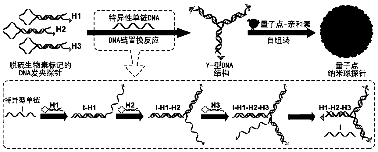
[0066] Quantum dot nanoball probes include streptavidin-coupled quantum dots, specific single-stranded DNA, and DNA hairpin probe 1, DNA hairpin probe 2 and DNA hairpin probe 3.

[0067] The specific single-stranded DNA contains 24 bases, and the specific sequence is as follows: 5'-GCACTA(y*)CTCCCT(b*)AACATC(x*)TCAAGC(a*)-3'.

[0068] DNA hairpin probe 1 contains 75 bases, and the specific sequence is as follows: 5'-AGGTTA(z)GCTTGA(a)GATGTT(x)AGGGAG(b)TAGTGC(y)TCCAAT(z*)CACAAC(c* ) GCACTA (y*) CTCCCT (b*) AACATC (x*) AAAAAAAAAAAAAAA-desthiobiotin-3'. Among them, z-a is a single chain, x-b-y and y*-b*-x* form a neck structure, z*-c* is a ring structure, A15 is a spacer arm, and desthiobiotin is a desthiobiotin group.

[0069] DNA hairpin probe 2 contains 75 bases, and the specific sequence is as follows: 5'-GATGTT(x)AGGGAG(b)TAGTGC(y)GTTGTG(c)ATTGGA(z)AACATC(x*)TCAAGC(a* ) TCCAAT (z*) CACAAC (c*) GCACTA (y*) AAAAAAAAAAAAAAA-de...

Embodiment 2

[0075] Embodiment 2 The preparation method of quantum dot nanoball probe

[0076] 1) Preparation of DNA hairpin probe solution: Dissolve the three synthetic desthiobiotin-modified DNA probe dry powders in ultrapure water to prepare 10 μM concentrated solutions, and store them at -20°C; 2 HPO 4 , 500mMNaCl) to dilute the probe concentrate into a 5μM solution; heat the prepared DNA probe solution in a 95°C water bath for 5min, then turn off the water bath heater, and slowly cool down to room temperature, and the DNA probe forming a hairpin structure is stored in 4°C.

[0077]2) Preparation of Y-shaped DNA structure: Mix 10 μL of the three DNA hairpin probes mentioned above, 10 μL of specific single-stranded DNA at a concentration of 5 μM, and 10 μL of phosphate buffer, and react at room temperature for 2 hours to form a Y-shaped structure. DNA structure.

[0078] 3) Preparation of quantum dot nanoball probes: fully mix the Y-shaped DNA structure solution with streptavidin-mod...

Embodiment 3

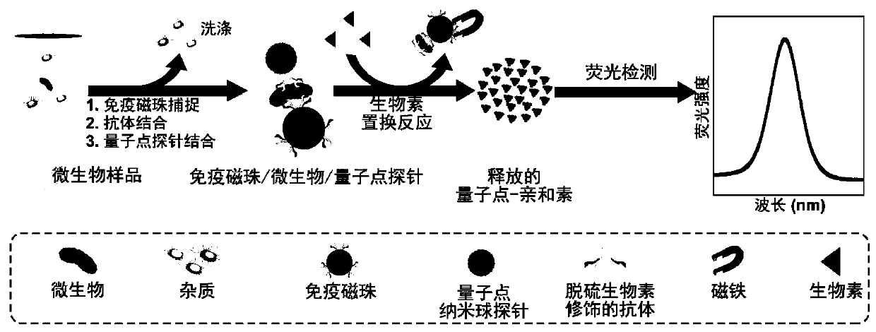
[0083] Embodiment 3 Rapid ultrasensitive detection method based on nanosphere probe

[0084] 1) Add the magnetic beads labeled with antibody A to the sample solution, and shake at room temperature (18-27°C) for 60 minutes to fully combine the antibody A on the magnetic beads with the target analyte; For immunomagnetic beads;

[0085] 2) Using a magnetic stand to separate the immunomagnetic bead-target analyte complex from the sample matrix, remove the sample matrix, and obtain the immunomagnetic bead-target analyte immune complex;

[0086] 3) Add desthiobiotin-modified specific antibody B to the immunomagnetic bead-target analyte complex obtained in 2), and shake at room temperature (18-27°C) for 60 minutes;

[0087] 4) Using a magnetic stand to separate the immunomagnetic bead-target analyte-antibody B complex from the unbound antibody to obtain the immunomagnetic bead-target analyte-antibody B immune complex;

[0088] 5) Add quantum dot nanoball probes to the immunomagneti...


PUM

PropertyMeasurementUnit
particle sizeaaaaaaaaaa

Abstract

The invention discloses a highly sensitive and decomposable quantum dot nanosphere probe and a preparation method thereof. Through a fulcrum-mediated strand displacement reaction, a DNA hairpin probe modified by three desulfated biotins is self-assembled into a Y-type DNA nanostructure; and the Y-type DNA structure and quantum dots coupled to streptavidin are self-assembled to form the quantum dot nanosphere probe. The prepared quantum dot nanosphere probe and an immunomagnetic separation technology are combined to be applied to quick detection of a target analyte. Because of a binding force between biotin and streptavidin stronger thana binding force of desulfurized biotin, the quantum dot nanosphere probe is decomposed, and thus quantum dots are released; the target analyte concentration is determined by detecting the fluorescence value of a quantum dot solution, and a shielding effect of immunomagnetic beads on the quantum dots is overcome. The method has high sensitivity and the detection limit of microorganisms is 1.37 cfu / mL. This method is good in specificity, and common other microorganisms do not affect the detection.

Description

technical field [0001] The invention belongs to the technical field of biological fluorescent probes, in particular to a highly sensitive and decomposable quantum dot nanoball probe and a preparation method thereof. Background technique [0002] Quantum dots are a kind of semiconducting nanomaterials, which have the advantages of long fluorescence lifetime, tunable emission wavelength and resistance to photobleaching. Bioluminescent probes prepared by coupling quantum dots with biomolecules such as antibodies and nucleic acids are widely used in medical diagnosis, food safety and environmental monitoring. For traditional quantum dot probes, one biorecognition molecule can only be coupled to one quantum dot particle, which has limited signal amplification and low detection sensitivity. Increasing the number of biorecognition molecules coupled with quantum dots is the key to preparing efficient fluorescent probes. Quantum dot-labeled DNA sequences can be assembled to form mu...

Claims


Application Information

Patent Timeline
Patent Type & Authority Patents(China)
IPC IPC(8): G01N33/533G01N33/543G01N33/58
Inventor 温俊林周顺桂余震陈俊华汤佳杨贵芹
Owner 天泓(济南)智能装备产业研究有限公司
Why Eureka
Try Eureka