Preparation method of bionic mineralized collagen scaffold

A technology of biomimetic mineralization and collagen scaffolds, applied in the field of preparation of biomimetic mineralization collagen scaffolds, can solve the problems of limited supply and high price

Active Publication Date: 2018-04-20
博纳格科技(天津)有限公司
View PDF5 Cites 17 Cited by
  • Summary
  • Abstract
  • Description
  • Claims
  • Application Information

AI Technical Summary

Problems solved by technology

[0005] Non-collagenous proteins play an important role in the biomineralization process, play an important regulatory role in stabilizing precursors and inducing the nucleation and hierarchical self-assembly of mineral salts within the collagen scaffold, but their supply is limited and expensive

Method used


Image

  • Preparation method of bionic mineralized collagen scaffold
  • Preparation method of bionic mineralized collagen scaffold
  • Preparation method of bionic mineralized collagen scaffold

Examples


Embodiment 1

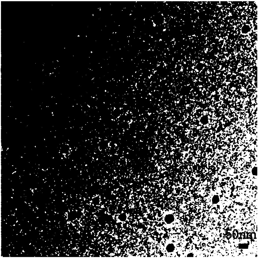
[0053] (1) Under the full stirring of a magnetic stirrer at room temperature, add 200 mg of O-carboxymethyl chitosan (viscosity: 45, Mw: 13700, isoelectric point 3.6) to 30 ml of deionized water, and after it is completely dissolved, add 41.76 mg K 2 HPO 4 , to obtain A solution; add 58.8mg CaCl in 10ml deionized water 2 2H 2 0, form B liquid; Under the sufficient stirring of normal temperature and magnetic stirrer, B liquid is added dropwise in A liquid, form carboxymethyl chitosan-amorphous calcium phosphate nanocomposite (CMC / ACP) solution; The pH of the CMC / ACP solution was adjusted to 2 with HCl. The structures characterized by transmission electron microscopy are as figure 2 As shown, the size of amorphous calcium phosphate nanoparticles is about 40-100 nm. SAED appears as a characteristic ring without concentric circles.

[0054] (2) Soak the dialysis bag in 50% alcohol by volume for 30 minutes, rinse with deionized water, seal one end, take 1ml type I rat tail c...

Embodiment 2

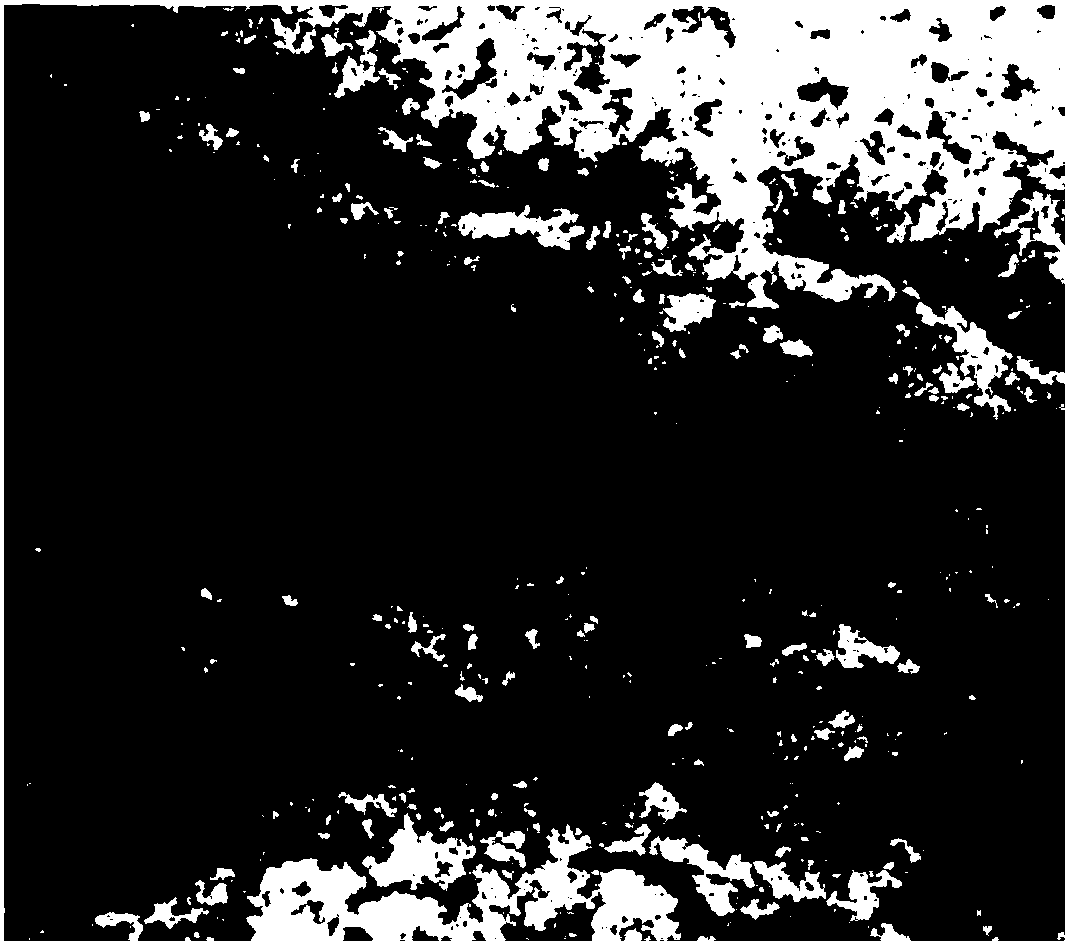
[0056] Under the sufficient stirring of the magnetic stirrer at room temperature, 200 mg of O-carboxymethyl chitosan (viscosity: 45, Mw: 13700, isoelectric point 3.6) added to 30 ml of deionized water, after completely dissolving, add 62.7 mg of K 2 HPO 4 , to obtain A solution; add 160.2mg SrCl in 10ml deionized water 2 ·6H 2 O, form B liquid; Under the sufficient stirring of normal temperature and magnetic stirrer, B liquid is added dropwise in A liquid, form carboxymethyl chitosan-amorphous strontium phosphate nanocomposite (CMC / ASP) solution; The pH of the CMC / ASP solution was adjusted to 2 with HCl. The structures characterized by transmission electron microscopy are as Figure 8 As shown, the size of CMC / ASP nanoparticles is about 5-50nm.

[0057] Soak the dialysis bag in 50% alcohol for 30 minutes, rinse with deionized water, seal one end, take 1ml type I rat tail collagen into the dialysis bag, take 3ml of the above acidic CMC / ASP solution, put it in the dialysis b...

Embodiment 3

[0059] Under the sufficient stirring of a magnetic stirrer at room temperature, 200 mg of O-carboxymethyl chitosan (viscosity: 45, Mw: 13700, isoelectric point 3.6) added to 30 ml of deionized water, after being completely dissolved, 25.44 mg of Na 2 CO 3 , to obtain A solution; add 106mgSrCl in 10ml deionized water 2 ·6H 2 O, form B liquid; Under normal temperature and sufficient stirring of a magnetic stirrer, B liquid is added dropwise in A liquid, form carboxymethyl chitosan-amorphous strontium carbonate nanocomposite (CMC / ASC) solution; The pH of the CMC / ASC solution was adjusted to 2 with HCl.

[0060] Soak the dialysis bag in 50% alcohol for 30 minutes, rinse with deionized water, seal one end, take 1ml type I rat tail collagen into the dialysis bag, take 3ml of the above acidic CMC / ASP solution, put it in a dialysis bag with a molecular weight cut off of 8000-14000 ,well mixed. After sealing, put 15ml of PBS buffer and mineralize for 3 days. The characterization re...


PUM

PropertyMeasurementUnit
particle diameteraaaaaaaaaa

Abstract

The invention provides a preparation method of a bionic mineralized collagen scaffold. The preparation method comprises the following steps: 1), preparing a bionic mineralized liquid: adjusting the pHvalue of carboxymethyl chitosan-amorphous calcium phosphate (or amorphous strontium phosphate or amorphous strontium carbonate) nanocomposite liquid to be lower than an isoelectric point of CMC; 2),preparing the bionic mineralized collagen scaffold: mixing the bionic mineralized liquid and acid-dissolved type-I collagen, then putting into a dialysis bag, sealing, then putting into a PBS buffer liquid, self-assembling and mineralizing for 2-5 days, then adding an alkaline CMC/ACP (ASP or ASC) liquid into the dialysis bag, and changing the PBS buffer liquid; 1-3 days later, putting the dialysis bag into deionized water for dialyzing for 10-72h, changing the deionized water for several times, centrifuging the liquid in the bag to obtain collagen gel, and performing freeze drying to obtain the bionic mineralized collagen scaffold. By the preparation method, collagen self-assembly and mineralization are cooperatively performed, intra-collagen fiber mineralization is achieved at relativelyhigh efficiency, and osteogenic ability-promoting strontium is integrated into the collagen scaffold, so that the preparation method has a wide clinical application prospect.

Description

technical field [0001] The invention belongs to the technical field of biomedical materials, and in particular relates to a preparation method of a bionic mineralized collagen scaffold. Background technique [0002] With the improvement of economic level and the maturity of implant restoration technology, implant restoration has the advantages of not harming adjacent teeth, beautiful and comfortable, so it has been widely used in dental clinics. The bone condition of the implanted area is of great significance to the osseointegration of the implant. Therefore, the treatment of the extraction socket after tooth extraction due to tooth and periodontal diseases is very important. In order to prevent and reduce the absorption of alveolar bone, bone tissue repair materials can be placed in the extraction socket for site preservation. Facial cysts and dead space left after tumor resection are often the main reasons for delayed wound healing. The placement of bone tissue repair ma...

Claims


Application Information

Patent Timeline
Patent Type & Authority Applications(China)
IPC IPC(8): A61L27/46A61L27/44A61L27/56
CPCA61L27/446A61L27/46A61L27/56A61L2430/02C08L89/00
Inventor 张旭林明丽沈敏娟刘欢欢王浩荣李彦秋
Owner 博纳格科技(天津)有限公司
Why Eureka
Try Eureka