A kind of lipid droplet fluorescent probe and its synthesis method and application

A technology of fluorescent probes and lipid droplets, applied in the direction of fluorescence/phosphorescence, chemical instruments and methods, luminescent materials, etc., can solve the problems of fewer fluorescent probes, high cost, cumbersome synthesis steps, etc., and achieve easy application and simple synthesis path , to achieve the effect of intracellular imaging

Inactive Publication Date: 2019-04-16
UNIV OF JINAN
View PDF3 Cites 0 Cited by
  • Summary
  • Abstract
  • Description
  • Claims
  • Application Information

AI Technical Summary

Problems solved by technology

[0005] Aiming at the problems in the prior art that there are few fluorescent probes for locating lipid droplets, cumbersome synthesis steps, and high cost, the present invention provides a lipid droplet fluorescent probe with two-photon properties that is easy to synthesize and accurate in positioning

Method used


Image

  • A kind of lipid droplet fluorescent probe and its synthesis method and application
  • A kind of lipid droplet fluorescent probe and its synthesis method and application
  • A kind of lipid droplet fluorescent probe and its synthesis method and application

Examples


Embodiment 1

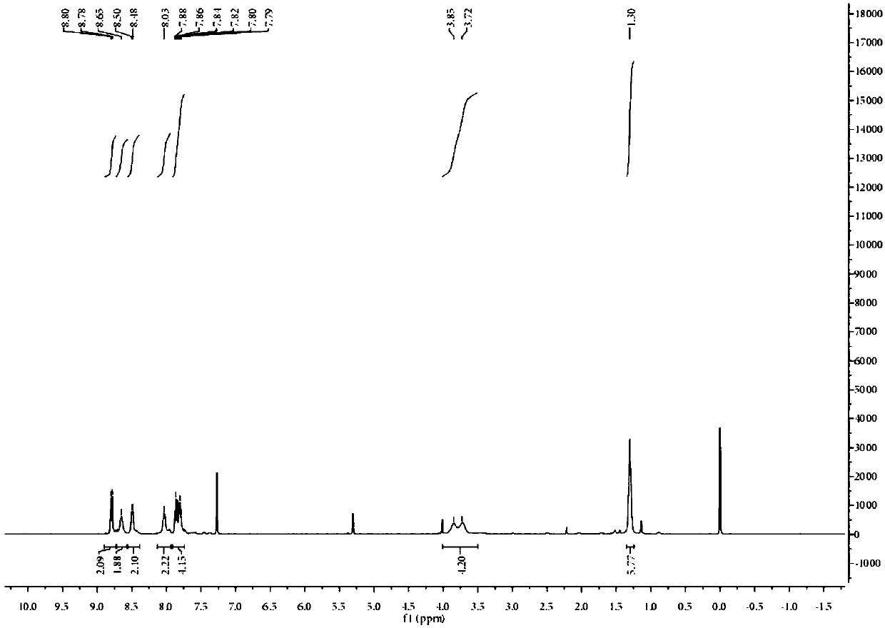
[0029] Example 1 Synthesis of PIA.

[0030] Dissolve 0.25g (1.2mmol) 9,10-phenanthrenequinone and 0.18g (1mmol) N,N-diethyl-4-aminobenzaldehyde in 20mL acetic acid, add 0.38g (5mmol) ammonium acetate as catalyst, The reaction was refluxed at 100 °C for 12 h under the protection of nitrogen. After the reaction, cool to room temperature, pour into ice water and stir, adjust the pH to neutral, filter with suction, and dry in a vacuum oven to obtain the probe compound with a yield of 80%. The proton magnetic spectrum of the probe compound is as follows figure 1 shown.

Embodiment 2

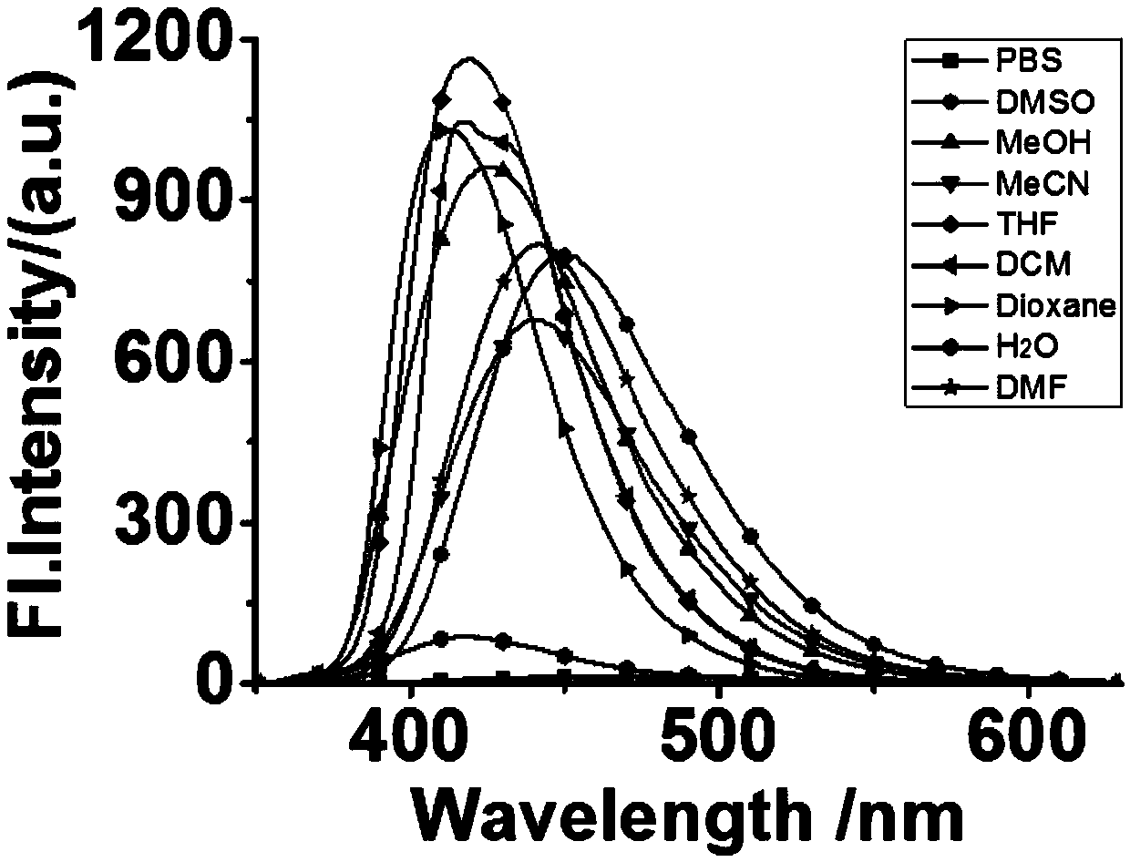
[0031] Example 2 Solvation effect of PIA.

[0032] The probe in Example 1 is dissolved in dimethyl sulfoxide, and it is prepared that the concentration is 10 -3 Mother liquor of M. Take 50 μL of the above mother solution and add it to nine identical 5mL volumetric flasks, and use DMF, DMSO, THF, MeCN, PBS, MeOH, DCM, H 2 O, Dioxane was diluted to 5mL, and then carried out fluorescence detection, the results are shown in figure 2 :Depend on figure 2 It can be seen that the fluorescence intensity of the probe in the organic solution is much greater than that in the aqueous phase. This shows that when the detection environment is the water phase, the fluorescence intensity of the probe in the water is obviously weaker, so that the probe can be better used in the detection of the human body environment, greatly reducing the harmfulness of the probe to the human body, and also eliminating self-disruptive.

Embodiment 3

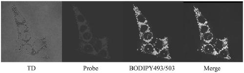
[0033] Example 3 Cellular colocalization of PIA.

[0034] The probe in Example 1 was made into a 1 mM stock solution with DMSO, and 5 μL of the stock solution was diluted with 1 mL of medium during staining to prepare a staining solution with a final concentration of 5 μM. The inoculated cells were incubated in the staining solution at 37 ºC for 30 min, washed 3 times with PBS, and the adherent cells were placed on a glass slide; then fluorescence imaging was performed with a fluorescence microscope, the excitation wavelength was 405nm, and the emission band was 425- 475nm. At the same time, co-localization experiments were carried out with a commercial lipid droplet probe (BODIPY493 / 503), with an excitation wavelength of 488 nm and an emission band of 500-550 nm. Images under a confocal fluorescence microscope as image 3 As shown, from left to right are bright field imaging, PIA probe imaging, BODIPY493 / 503 probe imaging, bright field, PIA probe and BODIPY493 / 503 probe sup...


PUM


Abstract

The invention provides a lipid droplet fluorescent probe, acquired by subjecting 9,10-phenanthraquinone and N,N-diethyl-4-aminobenzaldehyde to reflow reaction in acetic acid under ammonium acetate catalysis and protective gas, and separating. The lipid droplet fluorescent probe can detect lipid droplets by single-photon or dual-photon fluorescence during cell or tissue imaging.

Description

technical field [0001] The invention belongs to the field of organic small molecule fluorescent probes, in particular to a lipid drop fluorescent probe, in particular to a phenanthrenequinone compound fluorescent probe with two-photon properties. Background technique [0002] Lipid droplets are the main storage place for neutral lipids in cells, and they are widely found in bacteria, yeast, plants, insects and animal cells. Lipid droplets are spherical organelles whose main function is to dynamically regulate the energy balance of cells. In white adipose tissue, lipid droplets mainly store triglycerides and store energy; in brown adipose tissue, lipid droplets and mitochondria are closely connected to decompose triglycerides and provide energy for cells and biological organisms. Lipid droplets vary widely in size, ranging from 40 nm to 100 μm in diameter. Lipid droplets are composed of a monolayer of phospholipids and a hydrophobic core composed of neutral lipids, and ther...

Claims


Application Information

Patent Timeline
Patent Type & Authority Patents(China)
IPC IPC(8): C09K11/06C07D235/02G01N21/64
CPCC07D235/02C09K11/06C09K2211/1007C09K2211/1044G01N21/6486
Inventor 林伟英牛杰刘勇王伟珊
Owner UNIV OF JINAN
Why Eureka
Try Eureka