Fetal trophoblast cell separation method and application

A technology of trophoblasts and fetuses, applied in the medical field, can solve the problems of cumbersome steps, lack of economical practicability, uneconomical, etc., and achieve the effect of extensive clinical promotion value, extensive economical practicability, and simple cost

Pending Publication Date: 2018-08-17
宋清 +1
View PDF2 Cites 3 Cited by
  • Summary
  • Abstract
  • Description
  • Claims
  • Application Information

AI Technical Summary

Problems solved by technology

[0004] However, this method has limitations: First, the number of genetic diseases that can be detected by this method is very limited, only three; genetic diseases that cannot be detected, such as abnormalities in single gene diseases and polygenic diseases, and some fetal genomes Subtle changes in the fetus, especially some genetic abnormalities that lead to serious diseases or developmental disorders; second, the time point of making the diagnosis is late, and it has reached the mid-term induction period. mother's health
At present, the methods for isolating trophoblasts mainly use the specific markers of trophoblasts to activate cell sorting (Magnetically-activated cell sorting, MACS), fluorescence-activated cell sorting (Fluorescence-activated cell sorting, FACS) method, reference : [Durrant LG1, Martin WL, McDowall KM, Liu DT. Isolation of fetal trophoblasts and nucleated erythrocytes from the peripheral blood of pregnant women for prenatal diagnosis of fetal aneuploides. Early Hum Dev.1996 Dec 30; 47Suppl: S79-83], [ JF1, Metezeau P, Garcia-Fonknechten N, Richard Y, Tricottet V, Hsi BL, Kitzis A, Julien C, Papiernik E. Prenat Diagn. 1991 Oct; 11(10):787-98. Trophoblast-like cells sorted from peripheral maternal blood using flow cytometry: a multiparametric study involving transmission electron microscopy and fetal DNA amplification], this type of method is cumbersome, uneconomical, and requires certain equipment
In addition, some people use trophoblast size filtration to achieve the purpose of enrichment, and then still need the specific markers of trophoblasts or the separation method of laser microscopic capture to achieve the separation effect. References: [Mouawia H1, Saker A, Jais JP, Benachi A, Bussières L, Lacour B, Bonnefont JP, Frydman R, Simpson JL, Paterlini-Brechot P. Circulating trophoblastic cells provide genetic diagnosis in 63 fetuses at risk for cystic fibrosis or spinal muscular atrophy. Reprod Biomed Online. 2012 Nov. 25; 508-20], so the current method for isolating fetal trophoblast cells has the defects of cumbersome operation and lack of economical practicability,

Method used


Image

  • Fetal trophoblast cell separation method and application

Examples


Embodiment 1

[0039] Example 1 Analysis of Genomic DNA

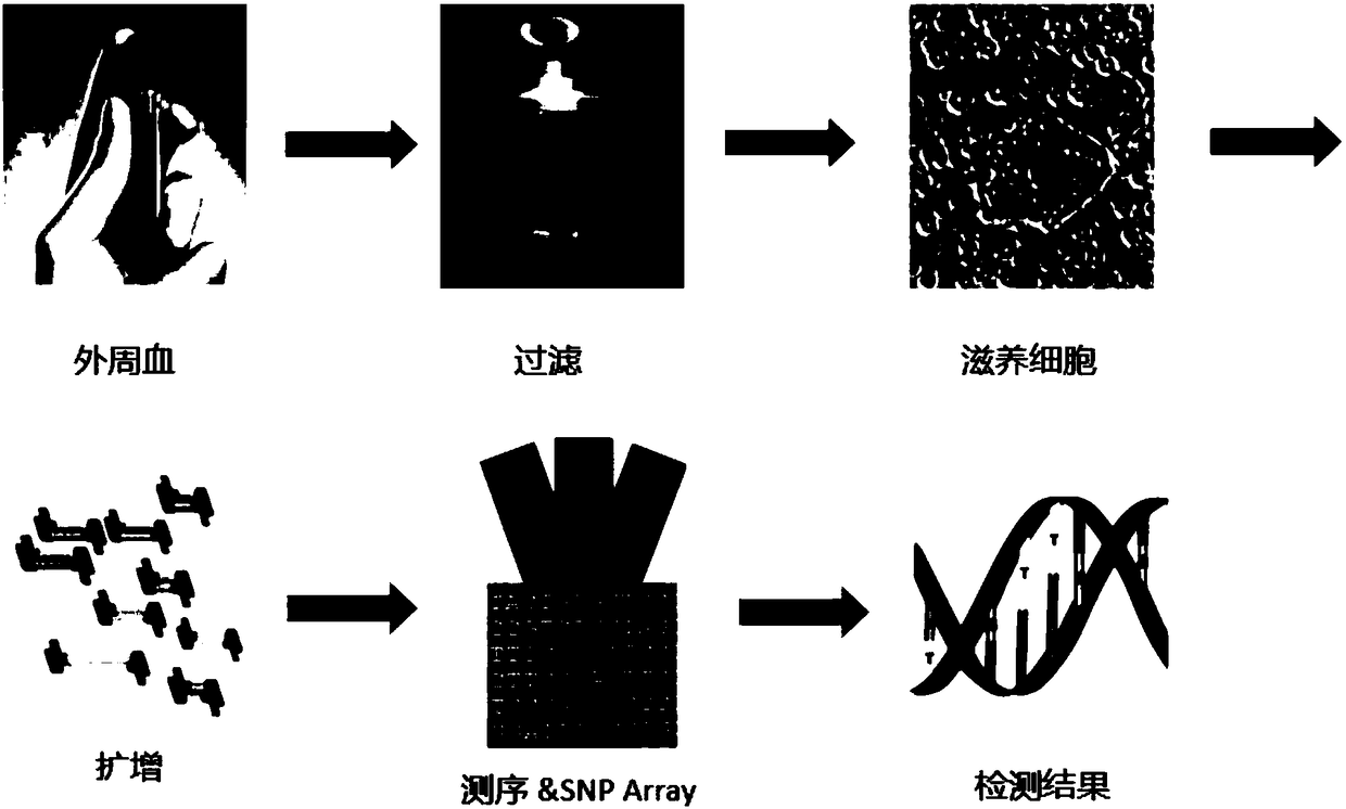
[0040] 1) Isolation of fetal trophoblast cells from maternal peripheral blood

[0041] Take 1-20ml of peripheral venous blood from pregnant women at 5-38 weeks of pregnancy, preferably at 8-15 weeks of pregnancy, the optimal blood collection volume is 10ml, and collect 10ml of blood into EDTA anticoagulant tubes (product number: 366643, BD); Within 1 hour, add 10ml of blood to Proprietary Buffer (Rarecells) and dilute it at 1:10. This Buffer can lyse red blood cells and fix nucleated cells; S1014) or filter with Chemrus disposable filter funnel (Chemrus, product number: CR-1008-300, 40μm) to remove non-trophoblast cells; if there are still other cells under the microscope, such as lymphocytes, granulocytes, etc., the filter can be repeated many times, Until pure maternal peripheral blood fetal trophoblasts are obtained;

[0042] 2), cell lysis and DNA amplification

[0043] After collecting a single fetal cell or a small number of ...

Embodiment 2

[0048] Example 2 Analysis of RNA expression

[0049] 1) Isolation of fetal trophoblast cells from maternal peripheral blood

[0050]Take 1-20ml of peripheral venous blood from pregnant women at 5-38 weeks of pregnancy, preferably at 8-15 weeks of pregnancy, the optimal blood volume is 10ml, and collect 10ml of blood into EDTA anticoagulant tubes (product number: 366643, BD). Within 4 hours of blood collection, add 10ml of blood to Proprietary Buffer (Rarecells) and dilute it at 1:10. This Buffer can lyse red blood cells and fix nucleated cells. After 10 minutes, the blood sample was filtered through a filter device with a pore size of 35 μm (Molecular Biotechnology, product number: S1014) or a Chemrus disposable filter funnel (Chemrus, product number: CR-1008-300, 40 μm) to remove non-trophic cells. If there are still other cells under the microscope, such as lymphocytes, granulocytes, etc., the filtration can be repeated many times until pure maternal peripheral blood fetal ...

Embodiment 3

[0055] Example 3 Analysis of DNA methylation

[0056] 1) Isolation of fetal trophoblast cells from maternal peripheral blood

[0057] Take 1-20ml of peripheral venous blood from pregnant women at 5-38 weeks of pregnancy, preferably at 8-15 weeks of pregnancy, the optimal blood volume is 10ml, and collect 10ml of blood into EDTA anticoagulant tubes (product number: 366643, BD). Within 4 hours of blood collection, add 10ml of blood to Proprietary Buffer (Rarecells) and dilute it at 1:10. This Buffer can lyse red blood cells and fix nucleated cells. After 10 minutes, pass the blood sample through a filter device with a filter aperture of 35 μm (Molecular Biotechnology, product number: S1014) or filter it with a Chemrus disposable filter funnel (Chemrus, product number: CR-1008-300, 40 μm) to remove non-trophic cells. There are still other cells under the microscope, such as lymphocytes, granulocytes, etc., which can be repeatedly filtered until pure maternal peripheral blood fet...


PUM

PropertyMeasurementUnit
pore sizeaaaaaaaaaa

Abstract

The invention discloses a fetal trophoblast cell separation method. According to the characteristic of large size of trophoblast cells, non-trophoblast cells in small sizes are removed by means of single or repeated filtration to obtain pure trophoblast cells. In addition, the invention discloses application of the fetal trophoblast cell to preparation of prenatal disgnosis medicines or kits. After one or more fetal trophoblast cells are obtained by separation, cell lysis is performed to directly obtain DNA, RNA, proteins, small RNA, methylated DNA and RNA, mitochondrial DNA and the like of the trophoblast cells, so that fetal information can be obtained. Compared with a previous fetal whole-genome acquisition method, the fetal trophoblast cell separation method and application of the fetal trophoblast cells in prenatal disgnosis have advantages of simplicity, economy, convenience in operation, high economic practicality and high clinical popularization value.

Description

technical field [0001] The invention relates to a method for non-invasively obtaining fetal trophoblast cells and detecting DNA information of fetal cells for prenatal diagnosis, belonging to the medical field. Background technique [0002] Genetic mutations refer to more than 7,000 known rare disease-causing genetic mutations and even unknown mutations and common disease-causing mutations. The existing technology only treats trisomy 21 (ie Down syndrome), Edwards syndrome (T18), Patau syndrome (T13) prenatal diagnosis of chromosomal disorders. In addition to the abnormal number of chromosomes, the fetus also has a large number of other genetic diseases such as single-gene diseases and polygenic diseases. [0003] One of the technical solutions for fetal genetic information detection currently on the market is non-invasive prenatal diagnosis. References: 【Chiu RW, Chan KC, Gao Y, Lau VY, Zheng W, Leung TY, Foo CH, Xie B, Tsui NB, Lun FM, Zee BC, Lau TK, Cantor CR, Lo YM. N...

Claims


Application Information

Patent Timeline
Patent Type & Authority Applications(China)
IPC IPC(8): C12N5/073C12Q1/6883G01N33/68
CPCC12N5/0603C12N2509/00C12Q1/6883C12Q2600/154C12Q2600/156C12Q2600/158C12Q2600/178G01N33/6893G01N2800/36
Inventor 宋清马丽
Owner 宋清
Why Eureka
Try Eureka