Method for ultrasensitive detection of miRNA based on dual amplification SERS signal system

A signal system and sensitive detection technology, applied in Raman scattering, material excitation analysis, etc., can solve the problems of insufficient sensitivity and time-consuming miRNA detection, achieve high sensitivity, simple detection process, and overcome the detection of low-expression short-sequence nucleosides acid effect

Active Publication Date: 2018-09-14
SOUTH CHINA NORMAL UNIVERSITY
View PDF5 Cites 8 Cited by
  • Summary
  • Abstract
  • Description
  • Claims
  • Application Information

AI Technical Summary

Problems solved by technology

Through bimetallic signal enhancement and enzyme digestion cycle system to amplify the signal, the double amplification of the signal is realized, and the rapid and high-sensitivity quantitative analysis of miRNA in the sample is realized, which solves the problems of insufficient sensitivity and long time-consuming detection of miRNA in the existing technology

Method used


Image

  • Method for ultrasensitive detection of miRNA based on dual amplification SERS signal system
  • Method for ultrasensitive detection of miRNA based on dual amplification SERS signal system
  • Method for ultrasensitive detection of miRNA based on dual amplification SERS signal system

Examples


Embodiment 1

[0060] (1), preparation of gold nanoparticles

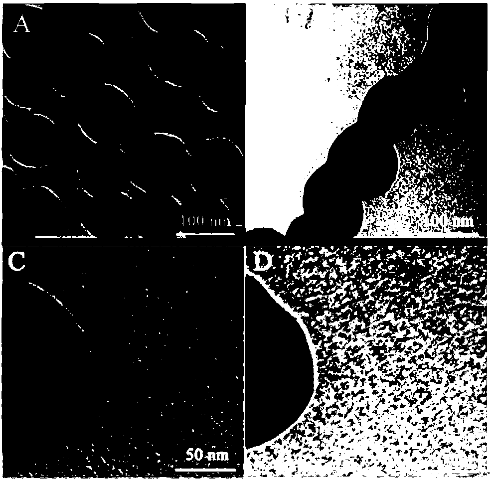
[0061] Add 20 mL of 1 mM ascorbic acid to 40 mL of 2 mM auric acid chloride (HAuCl) under constant stirring 4 )middle. The color change of the solution at this time is: light yellow-colorless-black-reddish brown, wait for the solution to turn reddish brown and continue to stir for 30 minutes. It was heated and boiled for 30 minutes, and finally cooled to room temperature to prepare gold nanoparticles with a diameter of 80 nm. figure 2 A is the TEM image of gold nanoparticles prepared under optimized conditions.

[0062] (2), preparation of polydopamine gold nanoparticles

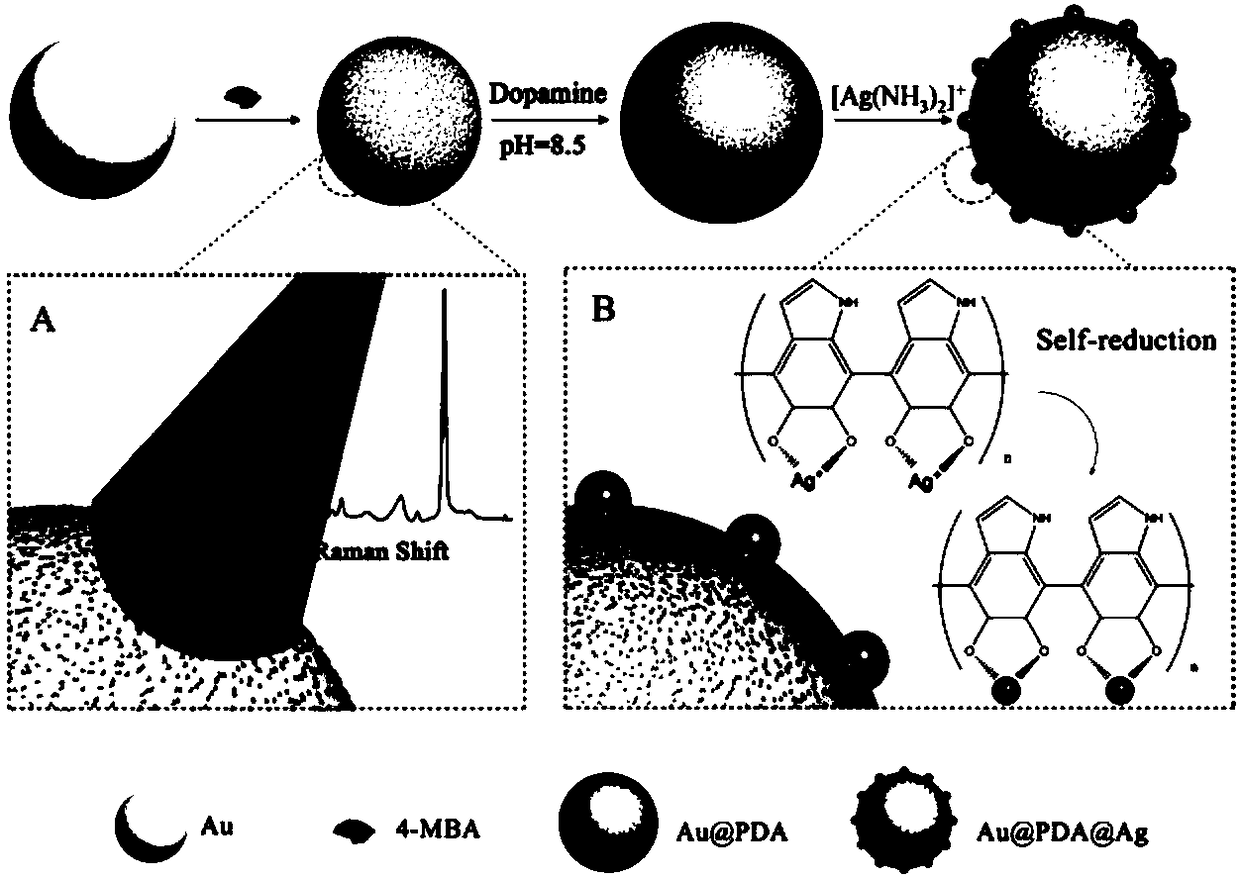
[0063] Take the above 1mL 1mmol / L gold nanoparticles, react with 50μL 100mM 4-MBA for about 30min, centrifuge (3000rpm, 10min) after the reaction, resuspend in Tris-HCl, add 200μL 0.02mg / mL dopamine hydrochloride alkaline solution, React at 37°C for 1 h, centrifuge (3000 rpm, 10 min) after the reaction, wash three times with triple distilled water, and res...

Embodiment 2

[0078] In order to illustrate the specificity of the present invention, two mutants of miRNA-31 were designed respectively, and the mutant sequences are as follows:

[0079] M1: 5'-AGGCAAGAUGTUGGCAUAGCU-3';

[0080] M2: 5'-AGGCAAGAUGCUTGCAUAGCU-3'.

[0081] The specific implementation steps refer to Example 1; the signal strength obtained can refer to Figure 7 , indicating that the present invention has strong specificity.

Embodiment 3

[0083] In order to test the application effect of this molecular diagnostic nanoplatform in real samples, we added miRNA-31 to human serum samples as a simulation of real samples. Prior to this, a series of treatments (filtration, centrifugation, dilution) should be performed on the serum to eliminate the possible influence of the matrix effect on the experimental results. Subsequently, different concentrations of miRNA-31 were added to the treated serum. These samples are respectively used for detection and analysis, and the specific implementation steps refer to Example 1; the obtained results are shown in Table 1, and the miRNA-31 content in healthy human serum is generally no more than 0.5fM, these results show that this method can It is very good for the detection of miRNA-31 in serum.

[0084] Table 1 Utilizes the molecular diagnostic nano-platform of the present invention to measure the results of miRNA-31 in simulated serum

[0085] miRNA-31(fM)


PUM

PropertyMeasurementUnit
particle sizeaaaaaaaaaa
particle diameteraaaaaaaaaa
wavelengthaaaaaaaaaa

Abstract

The present invention discloses a method for ultrasensitive detection of miRNA based on a dual amplification SERS signal system. The surface of 4-MBA-labeled gold nanoparticles is coated with a layerof polydopamine to form a SERS mark with a gold-PDA-silver satellite structure, the raman signal of the SERS mark is amplified greatly by unique localized surface plasmon resonance of gaps between gold and silver nanoparticles; at the same time, an enzymatic digestion circular reaction is combined with a magnetic capture mechanism to further amplify a SERS signal to achieve rapid and highly-sensitive quantitative detection of the miRNA. The method has the advantages of simple operation, good detection stability and high detection sensitivity; an incubation solution after the enzyme digestion circular reaction is sequentially added to functionalized magnetic nanoparticles for reaction, and then reacted with the SERS mark, and magnetic separation is performed just after the reaction is completed to achieve rapid and quantitative detection of the miRNA. The method can be applied to clinical diagnostic screening for early cancer.

Description

technical field [0001] The invention belongs to the technical field of biological detection, and in particular relates to a method for supersensitive detection of miRNA (microRNA) based on a double amplification SERS signal system. Background technique [0002] MicroRNA (miRNA) is a short-chain RNA in two types of non-coding (Non-coding) RNA, generally about 22 to 24 bases in size, widely involved in cell growth, proliferation and differentiation, and affects the physiological and pathological activities of the human body , and is closely related to the occurrence and development of tumors, which is a hotspot of current research. At present, the methods for detecting miRNA mainly include quantum dots, fluorescence method and qt-PCR, etc., but these methods have certain limitations due to their own characteristics, such as toxicity, easy photobleaching, complicated operation, etc., and cannot meet the measurement and detection requirements of modern medicine, etc. . However...

Claims


Application Information

Patent Timeline
Patent Type & Authority Applications(China)
IPC IPC(8): G01N21/65
CPCG01N21/65
Inventor 胡勇军江宁静朱挺锋余萌张文建王棋
Owner SOUTH CHINA NORMAL UNIVERSITY
Why Eureka
Try Eureka