Exosome-encapsulated nano drug-loading system for tumor treatment and preparation thereof
A nano-drug loading and tumor treatment technology, applied in the field of nanomaterials and oncology, can solve the problems of poor targeting and killing effect, reduce the proportion of CSCs, etc., and achieve the effect of a mild preparation method
- Summary
- Abstract
- Description
- Claims
- Application Information
AI Technical Summary
Problems solved by technology
Method used
Image
Examples
Embodiment 1
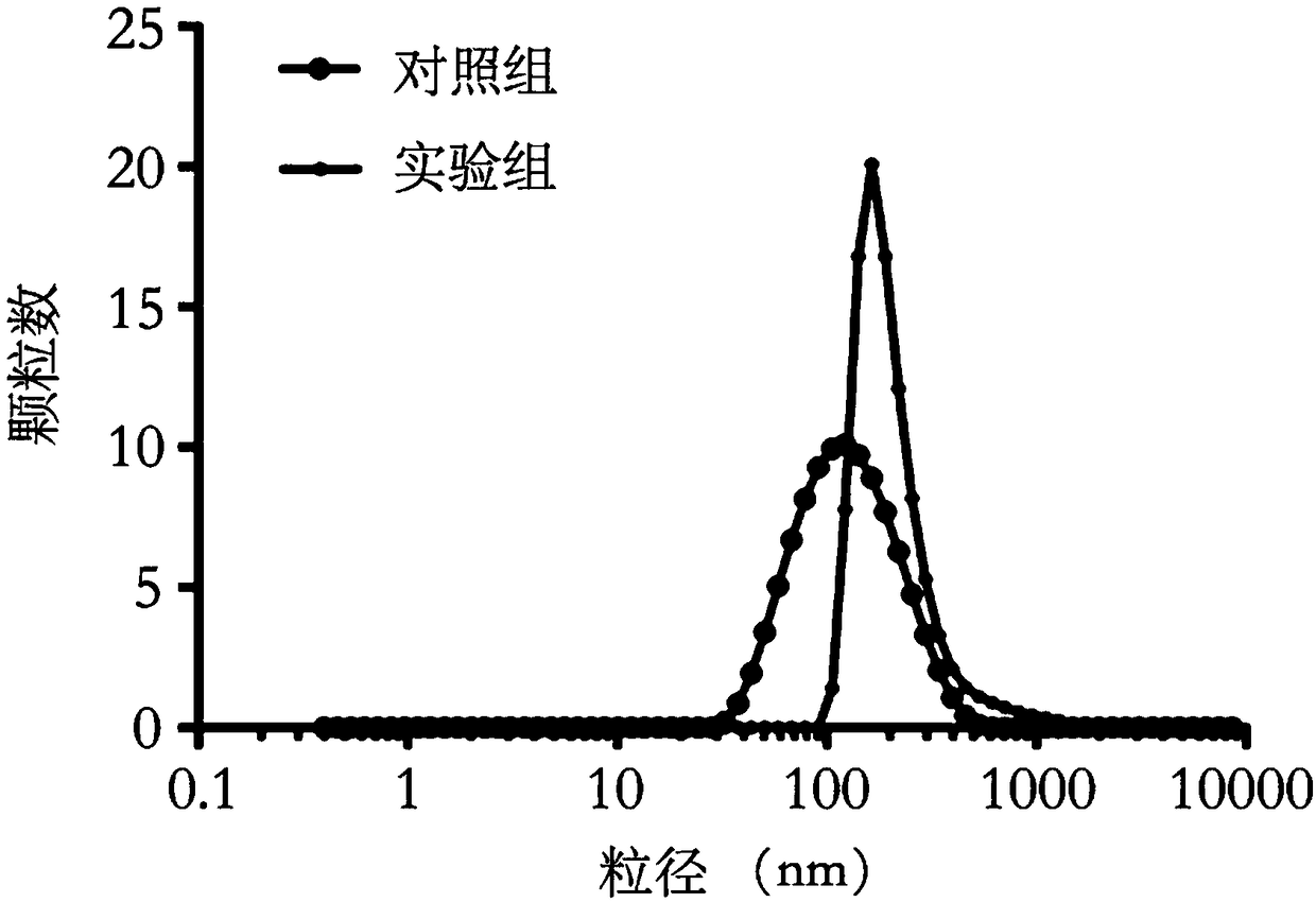
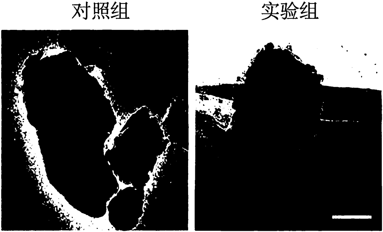
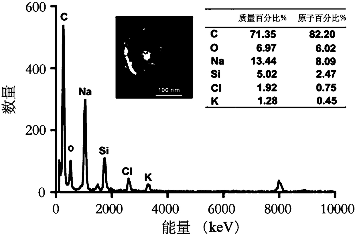
[0064] Example 1: Preparation and characterization of exosome-encapsulated nano-drug delivery system
[0065] 1. Experimental materials and reagents
[0066] H22 mouse liver cancer cells, doxorubicin (with red fluorescence), boron-doped p silicon wafer (resistance 0.00055-0.0015Ω-cm) were purchased from Virginia Company, USA
[0067] 2. Experimental steps
[0068] (1) Place the silicon wafer in concentrated H 2 SO 4 with H 2 o 2 in the mixed solution (volume ratio v:v=3:1) for 15 minutes, washed with ultrapure water for 3 times, and dried in the air for later use. Install the polished side of the silicon wafer in an electrochemical corrosion device, add HF / ethanol solution with a volume ratio of 4:1, 165mA / cm 2 After continuous corrosion for 300s at a certain current intensity, a dark red film appeared on the surface of the silicon wafer, washed with absolute ethanol for 3 times, and continued to add 3.3% HF / ethanol solution (mass ratio), 4.5mA / cm 2 Under the current in...
Embodiment 2
[0075] Example 2: Identification of surface membrane structure of exosome-encapsulated nano-drug delivery system
[0076] 1. Experimental materials and reagents
[0077] FITC-CD63 antibody, CD63 antibody, TSG101 antibody, Calnexin antibody
[0078] 2. Experimental steps
[0079] (1) Take a certain amount of E-PSiNPs, add 500 μL 5% BSA solution and incubate for 30 min, continue to add 10 μL FITC-CD63 antibody, incubate overnight at 4°C with shaking, centrifuge at 20,000g for 30 min, discard the supernatant, and wash 3 times with PBS. Take 20 μL of the E-PSiNPs solution incubated with FITC-CD63 and drop it on the confocal dish. After fully spreading, use the FV1000 confocal microscope to observe the co-localization of the green fluorescence of the FITC-CD63 antibody and the red fluorescence of PSiNPs. The specific parameters are: PSiNPs: Ex=488nm, Em=680nm; FITC: Ex=488nm, Em=520nm.
[0080] (2) Take E-PSiNPs, exosomes and corresponding cells with the same protein amount, and...
Embodiment 3
[0082] Example 3: Uptake behavior of exosome-encapsulated nano-drug delivery system in tumor cells and their CSCs in vitro
[0083] 1. Experimental materials and reagents
[0084] H22 mouse liver cancer cells, doxorubicin, 3D soft fibrin glue, 3D soft fibrin glue (1) In vitro tumor cell uptake behavior
[0085] 2×10 5 H22 was cultured in suspension in the cell culture plate. After 24 hours, the medium was removed, and 1 mL of serum-free DOX, DOX@PSiNPs or DOX@E-PSiNPs with DOX concentrations of 0.5, 1, and 2 μg / mL were added to the cell culture plate, respectively. Medium, 37°C, 5% CO 2 This normal cell culture condition was incubated for 4 hours, the cells were collected, washed 3 times with PBS, and centrifuged at 250g for 10 minutes. Add 500 μL of pre-cooled PBS to the cell pellet to resuspend the cells, pass through a 200-mesh sieve, and detect intracellular DOX fluorescence with a FC500 flow cytometer. Specific detection parameters: Ex=488nm, the emission light is det...
PUM
| Property | Measurement | Unit |
|---|---|---|
| Particle size | aaaaa | aaaaa |
Abstract
Description
Claims
Application Information
 - R&D
- Intellectual Property
- Life Sciences
- Materials
- Tech Scout
- Unparalleled Data Quality
- Higher Quality Content
- 60% Fewer Hallucinations
Browse by: Latest US Patents, China's latest patents, Technical Efficacy Thesaurus, Application Domain, Technology Topic, Popular Technical Reports.
© 2025 PatSnap. All rights reserved.Legal|Privacy policy|Modern Slavery Act Transparency Statement|Sitemap|About US| Contact US: help@patsnap.com


