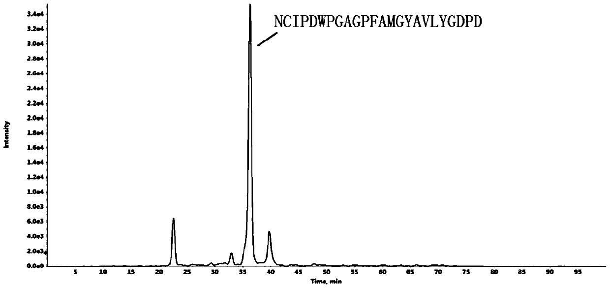
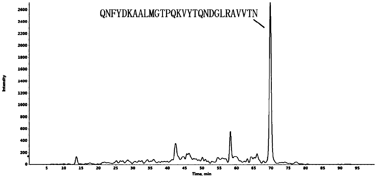
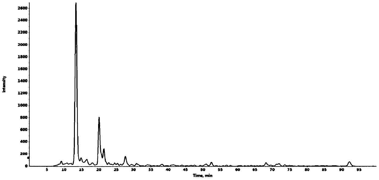
A method for identifying liver cancer stem cells
A liver cancer stem cell and identification method technology, applied in instruments, analytical materials, fermentation, etc., can solve the problems of low accuracy and cumbersome operation of detection methods, and achieve the effects of high accuracy, good application prospects, and convenient operation
- Summary
- Abstract
- Description
- Claims
- Application Information
AI Technical Summary
Problems solved by technology
Method used
Image
Examples
Embodiment 1
[0034] 1. Materials and methods
[0035] 1.1. Construction of LCSC cell lines
[0036] Take HepG2 cells, a human liver cancer cell line, and culture them in DMEM supplemented with 10% fetal bovine serum and 1% double antibody based on 5% CO 2 Incubate at 37°C in an incubator. HepG2 cells cultured to the exponential phase were digested with trypsin, and then treated with 20ng / mL basic fibroblast growth factor, 20ng / mL epidermal growth factor, 10ng / mL hepatic growth factor, b27, 1% methylcellulose Resuspended in DMEM / F12 medium with 1% double antibody, seeded the cell suspension into a low-adhesion 12-well plate at 2000 cells / mL, and placed in 5% CO 2 Incubate at 37°C in an incubator. Microcapsules with a diameter of about 200 μM are formed in 10 to 15 days. Obtain enriched cancer stem cell models.
[0037] Cells were sorted by flow cytometry using a flow cytometer. Liver cancer cells were first labeled with anti-CD133 monoclonal antibody, then washed with PBS and resuspende...
Embodiment 2
[0055] A method for identifying liver cancer stem cells, comprising the following steps:
[0056] 1) Take the MEM medium that has eliminated L-asparagine, L-aspartic acid and glycine as the cell culture medium, and in CO 2 In an incubator, culture the cell mass to be tested in the cell culture medium at a temperature of 28° C. in the dark for 16 hours;
[0057] 2) Prepare an aqueous solution containing the following components as an inducer: HuEPO 25 μg / L, polyoxyethylene lauryl ether 80 μg / L, polyvinylpyrrolidone 40 μg / L, chitosan 200 μg / L, sodium chloride 1 mg / L, p-hydroxyl Ethyl benzoate mg / L, BSA 150 μg / L, phosphate buffer 1 mg / L; take the product obtained in step 1), collect the cell mass, insert it into the inducer, and place it under anaerobic and light-proof conditions at a temperature of 35°C. Keep 24h under the condition;
[0058] 3) Take the product obtained in step 2), ultrasonically break it at a frequency of 15kHz for 8min, collect the product and centrifuge at...
Embodiment 3
[0061] A method for identifying liver cancer stem cells, comprising the following steps:
[0062] 1) Take the MEM medium that has eliminated L-asparagine, L-aspartic acid and glycine as the cell culture medium, and in CO 2 In an incubator, culture the cell mass to be tested in the cell culture medium at a temperature of 28° C. in the dark for 16 hours;
[0063] 2) Prepare an aqueous solution containing the following components as an inducer: HuEPO 25 μg / L, polyoxyethylene lauryl ether 80 μg / L, polyvinylpyrrolidone 40 μg / L, chitosan 200 μg / L, sodium chloride 1 mg / L, p-hydroxyl Ethyl benzoate mg / L, BSA 150 μg / L, phosphate buffer 1 mg / L; take the product obtained in step 1), collect the cell mass, insert it into the inducer, and place it under anaerobic and light-proof conditions at a temperature of 35°C. Keep 24h under the condition;
[0064] 3) Take the product obtained in step 2), ultrasonically break it at a frequency of 15kHz for 8min, collect the product and centrifuge at...
PUM
 Abstract
Description
Claims
Application Information
 - R&D
- Intellectual Property
- Life Sciences
- Materials
- Tech Scout
- Unparalleled Data Quality
- Higher Quality Content
- 60% Fewer Hallucinations
Browse by: Latest US Patents, China's latest patents, Technical Efficacy Thesaurus, Application Domain, Technology Topic, Popular Technical Reports.
© 2025 PatSnap. All rights reserved.Legal|Privacy policy|Modern Slavery Act Transparency Statement|Sitemap|About US| Contact US: help@patsnap.com


