Detection method for monomolecular protein

A detection method and single-molecule technology, applied in the field of protein detection, can solve the problems of inability to meet the detection requirements of proteins with low expression abundance and low detection sensitivity

Active Publication Date: 2019-01-04
PILOT GENE TECH HANGZHOU CO LTD
View PDF3 Cites 7 Cited by
  • Summary
  • Abstract
  • Description
  • Claims
  • Application Information

AI Technical Summary

Problems solved by technology

[0004] In view of this, the purpose of the present invention is to provide a single-molecule protein detection method for the problems of low detection sensitivity in the prior art and the inability to meet the detection requirements for proteins with low expression abundance

Method used


Image

  • Detection method for monomolecular protein
  • Detection method for monomolecular protein
  • Detection method for monomolecular protein

Examples


Embodiment 1

[0080] 1. Laser confocal detection system ( figure 1 with figure 2 )

[0081] The laser confocal detection system includes a microfluidic chip A, a point light source B, an imaging unit C, a scanning lens D and a three-dimensional droplet imaging unit.

[0082] The point light source B adopts a laser or an LED lamp to form a point light source and project it onto the droplet.

[0083] The imaging unit C is an EMCCD photon detector, and the imaging unit C collects and detects the fluorescent signals processed by the four-channel fluorescent filter F and the lens G, and can use different fluorescent channels to independently synthesize the characteristics of the droplet containing the fluorescent signal Numerical imaging of droplet shape and fluorescence intensity °C.

[0084] 2. Microfluidic chip

[0085] The preparation of the hot-pressing mold, using the nickel mold to prepare the upper structure, see the detailed structure figure 1, wherein the width of the droplet for...

Embodiment 2

[0094] Example 2: Detection of GPC1 by direct fluorescence method

[0095] Preparation of multilayer droplets:

[0096] (1) Prepare the oil phase. Prepare the oil phase according to the following proportions (both by mass): 50g mineral oil + 50g n-tetradecane + 1.5g emulsifier EM90 + 1.5g TritonX-100, and use it for later use after ultrasonic degassing.

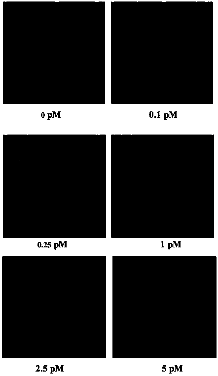
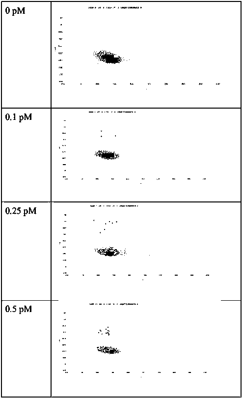
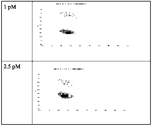
[0097] (2) Prepare the water phase. Prepare different concentrations of GPC1 protein standards (abcam, ab114484), that is, use the standard diluent to dilute the original GPC1 protein standard, and the final GPC1 protein concentrations are 0, 0.1, 0.25, 0.5, 1.0, 2.5, 5 and 10PM, respectively. , the final volume is not less than 200 μl. Use 200 μl of GPC1 protein in 6 μL capture solution of Merck’s magnetic bead European GPC1 capture antibody (abcam, ab55971), incubate at 37 ° C for 1 h, wash the supernatant, and then add 200 μl of FITC-labeled detection antibody (Invitrogen, Cat#62-8411) , the detection antibody was dilu...

Embodiment 3

[0101] Example 3: Detection of GPC1 by HRP enzyme labeling method

[0102] Preparation of multilayer droplets:

[0103] (1) Prepare the oil phase. Prepare the oil phase according to the following proportions (both by mass): 50g mineral oil + 50g n-tetradecane + 1.5g emulsifier EM90 + 1.5g TritonX-100, and use it for later use after ultrasonic degassing.

[0104] (2) Prepare the water phase. Prepare different concentrations of GPC1 protein standards (abcam, ab114484), that is, use the standard diluent to serially dilute the original GPC1 protein standard, to the final GPC1 protein concentration of 0, 0.1, 0.25, 0.5, 1.0, 2.5, 5 and 10pM The final volume is not less than 200 μl. 200 μl of GPC1 protein in 6 μl capture solution of Merck’s magnetic bead Eurolink GPC1 capture antibody (abcam, ab55971), incubated at 37°C for 1 h, washed the supernatant, and then added 200 μl of HRP-labeled detection antibody (Invitrogen, Cat#31430). The detection antibody was diluted with PBS at ...


PUM

PropertyMeasurementUnit
particle diameteraaaaaaaaaa
lengthaaaaaaaaaa

Abstract

The invention belongs to the field of protein detection and discloses a detection method for monomolecular protein. The detection method for the monomolecular protein comprises the following steps: mixing to-be-detected protein and a target protein antibody to generate a to-be-detected protein and target protein antibody compound, mixing the to-be-detected protein and target protein antibody compound and a detection antibody marked by a marker, performing dissociation and collecting a to-be-detected protein and detection antibody compound; directly dispersing the to-be-detected protein and detection antibody compound into a micro-fluidic chip, or mixing the to-be-detected protein and detection antibody compound and a substrate corresponding to an enzyme marker, carrying out a reaction anddispersing reaction liquid into the micro-fluidic chip; performing fluorescent signal scanning by a laser confocal detection system and performing monomolecule counting. According to the detection method, the monomolecular protein can be detected quantitatively and rapidly; the detection method is high in detection sensitivity and suitable for protein with too low expression abundance. The detection method is convenient to operate, accurate in detection result and small in CV value deviation; furthermore, the flux is high and multiple samples can be detected at a time.

Description

technical field [0001] The invention belongs to the field of protein detection, and in particular relates to a single-molecular protein detection method. Background technique [0002] Protein, as the most important component of the body, interprets the growth, development and change of every point of the individual life. Since the 1940s, with the development of classical protein detection techniques such as immunohistochemistry, the value of protein as a biomarker has gradually been valued by people. Although immunohistochemistry, ELISA, Luminex and other protein detection technologies have realized the detection of thousands of protein biomarkers, the development speed of protein biomarkers is still slow: only 1-2 new biomarkers per year into practical clinical applications. According to statistics, among more than 400,000 known human proteins, about 300,000 cannot be detected by traditional methods due to their low expression abundance. Of the approximately 100,000 prot...

Claims


Application Information

Patent Timeline
Patent Type & Authority Applications(China)
IPC IPC(8): G01N33/68G01N21/64
CPCG01N21/6428G01N33/68
Inventor 宋小慧位玉玲夏江
Owner PILOT GENE TECH HANGZHOU CO LTD
Why Eureka
Try Eureka