Fluorescence immunochromatographic kit for quantitative joint detection of four urinary micro proteins and preparation method thereof

A fluorescent immunochromatography and immunochromatography test paper technology, which is applied in measurement devices, analytical materials, biological tests, etc., can solve the problems of not being able to perform simultaneous detection, time-consuming and laborious, etc., and achieve fast and convenient detection, cost reduction, and simple operation process. Effect

Inactive Publication Date: 2019-04-12
杭州康知生物科技有限公司
View PDF9 Cites 11 Cited by
  • Summary
  • Abstract
  • Description
  • Claims
  • Application Information

AI Technical Summary

Problems solved by technology

[0005] The existing clinical detection of the above-mentioned trace proteins in urine is usually a single detection, which cannot be detected at the same time, which is time-consuming and labor-intensive

Method used


Image

  • Fluorescence immunochromatographic kit for quantitative joint detection of four urinary micro proteins and preparation method thereof
  • Fluorescence immunochromatographic kit for quantitative joint detection of four urinary micro proteins and preparation method thereof
  • Fluorescence immunochromatographic kit for quantitative joint detection of four urinary micro proteins and preparation method thereof

Examples


preparation example Construction

[0042] Preparation of Antigen Solution Labeled with Time-Resolved Fluorescent Microspheres

[0043] First, weigh 1.5 mg of EDC (1-(3-dimethylaminopropyl)-3-ethylcarbodiimide hydrochloride), add 150ul of boric acid buffer solution (0.05M, pH=8.0) and vortex to dissolve , prepared as a 10mg / ml solution;

[0044] Next, take four 1.5ml centrifuge tubes, add 40ul boric acid buffer solution (0.05M, pH=8.0) and 10ul time-resolved fluorescent microspheres respectively, vortex, mix well, and add 1.5ul to the four centrifuge tubes respectively EDC (10mg / ml) solution, activated by shaking at room temperature for 15min (note: mix immediately after adding EDC), centrifuge (15000rpm, 10°C, 10min), discard the supernatant, and resuspend with 40ul boric acid buffer solution (0.05M), Ultrasonic dispersion (100W, 1s*10);

[0045] Then, in the four centrifuge tubes, add the antigen and protein A according to the following amounts respectively, vortex and mix well, and place on a shaker at 4° C...

Embodiment 1

[0076] Embodiment 1 sensitivity test

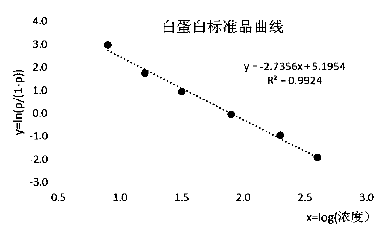
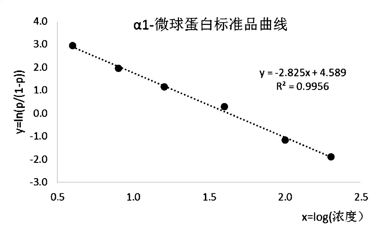
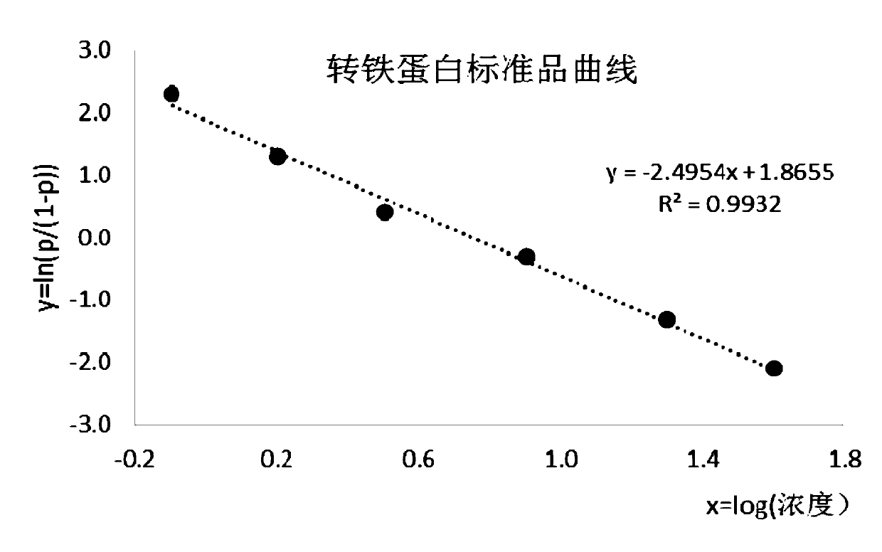
[0077] Mix and dilute the four antigen proteins labeled with fluorescent microspheres to the working concentration to obtain the antigen solution labeled with fluorescent microspheres (the working concentration is diluted according to the ratio of 1:1000-3000), take 20ul of 10mM PBS as a blank sample, and add it to the layer Analyze the test strips, and then add 80ul of mixed and diluted fluorescent microsphere-labeled antigen solution. After reacting for 15 minutes, use an immunofluorescence analyzer to read the four T-line and C-line fluorescence signals on the nitrocellulose membrane and calculate the ratio T / (T+C) respectively. Repeat the test 20 times. And calculate the mean M and standard deviation SD of 20 test results. Then calculate the value of M-2SD, the result is shown in the table below. Substitute the value of M-2SD into the fitted standard curve, and calculate the corresponding concentration value, which is the sensitivi...

Embodiment 2

[0082] Embodiment 2 precision test

[0083] Mix and dilute the antigenic proteins of four kinds of labeled fluorescent microspheres to the working concentration to obtain the antigen solution labeled with fluorescent microspheres (the working concentration is diluted according to the ratio of 1:1000-3000), and test with three samples of different concentrations. Take 20ul of the sample, add it to the chromatographic test strip, and then add 80ul of the diluted fluorescent microsphere-labeled antigen solution. After reacting for 15 minutes, use an immunofluorescence analyzer to read 4 T lines and C line fluorescence signals on the nitrocellulose membrane, and repeat the test 10 times. Calculate the ratio T / (T+C) respectively and substitute the ratio into the standard substance fitting curve equation, calculate its corresponding concentration value, and calculate its mean M and standard deviation SD. According to the formula coefficient of variation CV (%)=SD / M*100%, respective...


PUM

PropertyMeasurementUnit
Sensitivityaaaaaaaaaa

Abstract

The invention discloses a fluorescence immunochromatographic kit for quantitative joint detection of four urinary micro proteins and a preparation method thereof, which solves the problems that the detection of urinary micro proteins in clinical detection cannot be simultaneously carried out. A fluorescence immunochromatographic kit for quantitative joint detection of four urinary micro proteins comprises an immunochromatographic strip coated with an albumin capture antibody, an alpha 1-microglobulin capture antibody, a transferrin capture antibody, a beta 2-microglobulin capture antibody anda protein A, and a fluorescent microsphere labeled antigen solution including a fluorescent microsphere labeled albumin solution, a fluorescent microsphere labeled alpha 1-microglobulin solution, a fluorescent microsphere labeled transferrin solution and a fluorescent microsphere labeled beta 2-microglobulin solution; and fluorescent microspheres are also labeled with IgG immunoglobulin. The invention has the advantages of rapid detection and the like.

Description

technical field [0001] The invention belongs to the field of in vitro diagnostic immunological detection, in particular to a fluorescent immunochromatographic kit for quantitative joint detection of four trace proteins in urine and a preparation method thereof Background technique [0002] A urine test is a medical testing method. Including quantitative determination of protein composition, etc. Determination of microalbumin in urine can reflect early renal disease and renal damage. Elevated trace protein in urine is more common in the pre-stage of kidney disease complicated by diabetes, hypertension, pregnancy eclampsia and other diseases. The early detection stage of urinary microalbumin is an early signal and harbinger of nephropathy. At this time, the kidney damage is still in a reversible period. If treated in time, the development process of nephropathy can be terminated or reversed. [0003] The combined detection of urine microprotein can be used as an indicator o...

Claims


Application Information

Patent Timeline
Patent Type & Authority Applications(China)
IPC IPC(8): G01N33/68G01N33/533G01N33/543
CPCG01N33/533G01N33/54306G01N33/54313G01N33/6893
Inventor 张乐之吴敏华余铭恩王立童吴滨胡祥叶
Owner 杭州康知生物科技有限公司
Why Eureka
Try Eureka