
Gene mutation detection method based on selective elimination of wild strand background interference

A detection method and single-chain technology, applied in biochemical equipment and methods, microbial determination/inspection, etc., can solve the problems of high price of instruments and reagents, insufficient sensitivity, long detection period, etc.

Active Publication Date: 2019-04-26
PEKING UNIV
View PDF6 Cites 2 Cited by
  • Summary
  • Abstract
  • Description
  • Claims
  • Application Information

AI Technical Summary

Problems solved by technology

[0010] To sum up, the existing methods still have a series of problems in early tumor detection of ctDNA, such as insufficient sensitivity, long detection cycle, unstable methods, harsh experimental conditions and high prices of instruments and reagents.

Method used


Image

  • Gene mutation detection method based on selective elimination of wild strand background interference
  • Gene mutation detection method based on selective elimination of wild strand background interference
  • Gene mutation detection method based on selective elimination of wild strand background interference

Examples


Embodiment 1

[0052] Example 1

[0053] In this embodiment, two different target gene DNA sequences were selected, one is the sequence where the point mutation (L858R) at codon 858 of exon 21 of the human epidermal growth factor receptor (EGFR) is located; the other is the sequence of V - the sequence of the point mutation (G13D) at codon 13 of exon 2 of Ki-ras2Kirsten rat sarcoma virus oncogene (KRAS).

[0054] Table 1. Sequences (5'-3') of nucleic acid strands used in this example

[0055]

[0056]

[0057]

[0058] (1) EGFR L858R sulfurated DNA strand guides DNase I to selectively cut EGFR L858R wild-type DNA strand

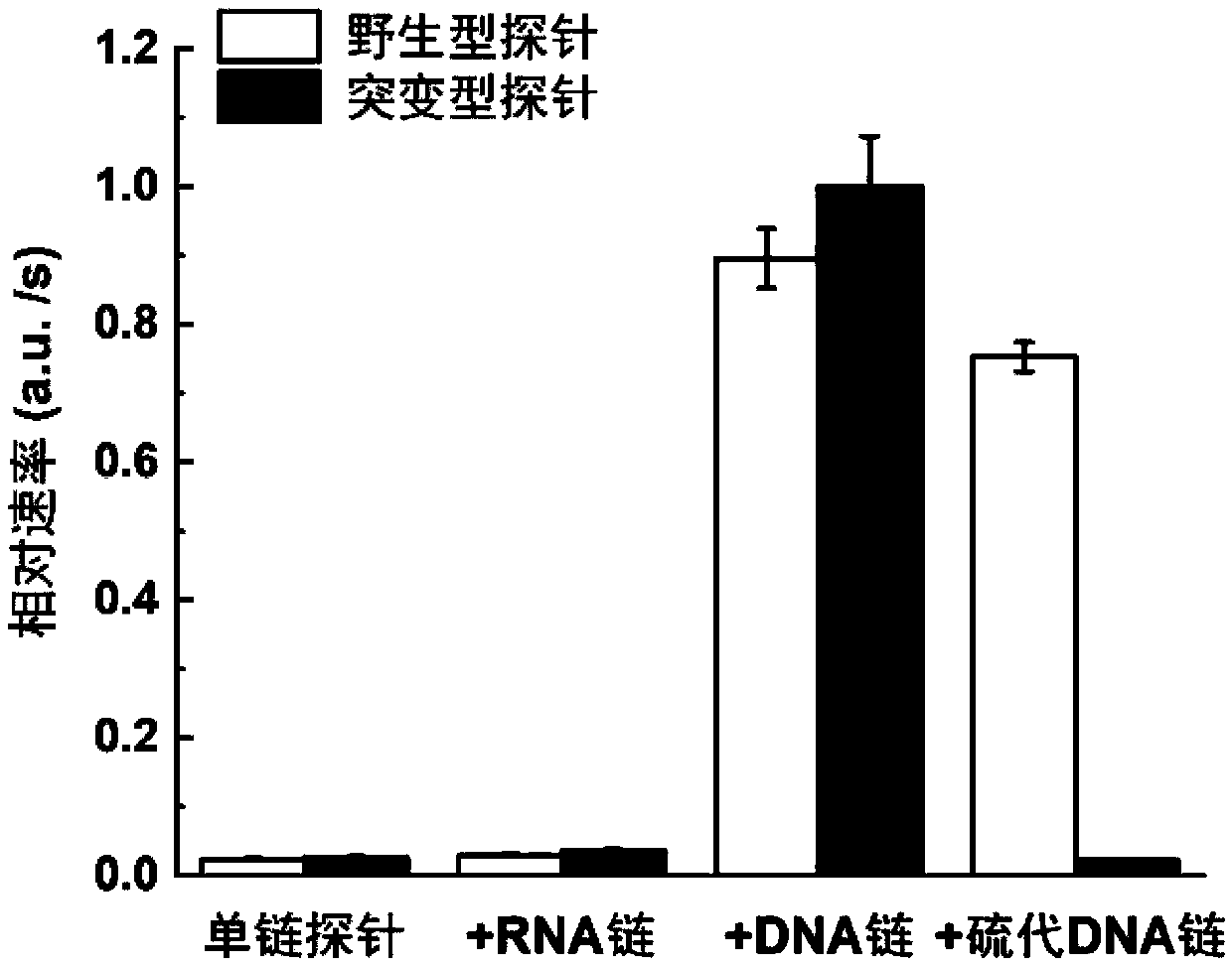
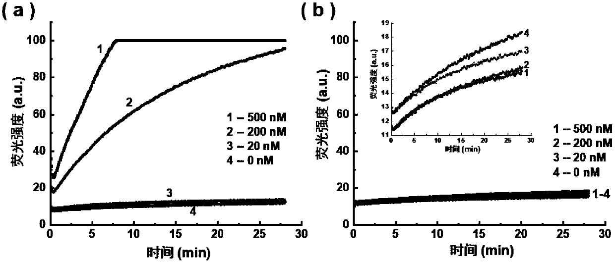
[0059] The sequences of the EGFR L858R wild-type long-chain DNA and the EGFRL858R mutant long-chain DNA are shown in Table 1, the underlined part is the target region, and the mutation site is indicated in bold. For this target region, design the corresponding EGFR L858R sulfide DNA strand sequence as: 5'-AAAAAAAAAAAA GGGCTGGCCAA CGCAGATA-3', wherein the phosp...

Embodiment 2

[0082] Example 2

[0083] The sequences of wild-type DNA long chain, mutant DNA long chain, thio-DNA chain and RNA closed chain of EGFR L858R used in this example are listed in Table 1. The sequences of the PCR amplification primers used are:

[0084] EGFR L858R forward primer: 5'-TTCTTTTCTCTTCCGCACC-3' (5'-OH end) (SEQ ID No: 21)

[0085] EGFR L858R phosphorylation reverse primer: 5'-PO 4 -TACTTGGAGGACCGTCG-3' (5' terminal phosphorylation tag) (SEQ ID No: 22)

[0086] The experimental operation steps are as follows:

[0087] 1) Genomic DNA was digested with Shearase enzyme: Mix 1 mg of genomic DNA and 1.5 μL Shearase enzyme in 20 μL buffer, the buffer composition was 10 mmol / L Tris-HCl, 25 mmol / L MgCl 2 , 1mmol / LDTT, pH 7.5@25°C, incubate at 42°C for 15min, then heat inactivate the Shearase enzyme.

[0088] 2) PCR amplification of genomic DNA and standard DNA: 1.5 μL of the product solution in step 1) or 0.25 amol DNA standard, 20 pmol forward primer, 20 pmol reverse pr...


PUM


Abstract

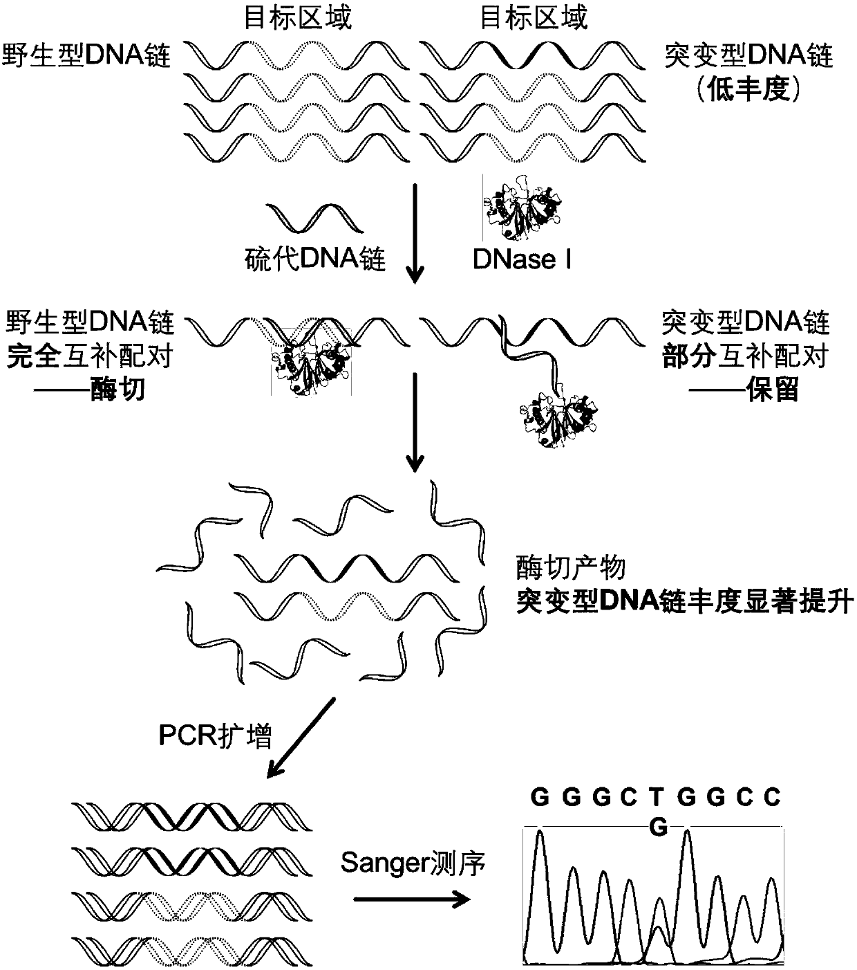
The invention discloses a gene mutation detection method based on selective elimination of wild strand background interference. A target sequence to be detected is amplified through PCR (Polymerase Chain Reaction) and then is processed into single-stranded DNA (Deoxyribonucleic Acid) through Lambda exonuclease; a thio-DNA strand which is complementary with a target region of a wild type DNA sequence and an RNA (Ribonucleic Acid) closed strand which is complementary with a non-target region are designed and synthesized; then the thio-DNA strand and the RNA closed strand are mixed with the single-stranded DNA, and DNase I is added to carry out cutting after temperature rising and annealing are carried out; a wild type DNA strand target region sequence is selectively cut off through the DNaseI under the guidance of the thio-DNA strand; a mutation type DNA strand is remained since mispairing exists in the target region and cannot be cut off by the DNase I, so that the abundance of the mutation type DNA strand is remarkably improved and the difficulty in detecting low-abundance gene mutation in the prior art is greatly reduced. The method disclosed by the invention does not need complicated and expensive instruments, is easy to operate and low in cost and can provides a result within 1 day; in-time and reliable gene mutation information can be provided for clinical early screeningof tumors and monitoring the recurrence after operation.

Description

technical field [0001] The present invention relates to the field of genome sample processing and gene mutation detection, and in particular to a method for pretreatment of genome samples. After the wild-type DNA is eliminated by this method, it is used in conjunction with conventional sequencing methods to realize rapid detection of ultra-low abundance gene mutations. , Low-cost sequencing analysis. Background technique [0002] Cancer (malignant tumor) is the number one killer that seriously threatens human health today, and early diagnosis and postoperative recurrence monitoring related to cancer have important biological and medical significance. Gene mutation refers to the heritable variation of genomic DNA molecules, which is one of the important reasons leading to the formation of malignant tumors. The traditional method of tumor gene detection is mainly tissue biopsy, that is, to obtain tumor tissue samples through surgical puncture and perform genetic detection on ...

Claims


Application Information

Patent Timeline
Patent Type & Authority Applications(China)
IPC IPC(8): C12Q1/6806
CPCC12Q1/6806C12Q2531/113C12Q2521/319C12Q2521/345
Inventor 赵美萍陈维阳彝栋肖先金李梦圆
Owner PEKING UNIV
Learn More