Pepsinogen I/pepsinogen II detection kit

A technology of pepsinogen and detection kits, applied in the direction of measuring devices, instruments, scientific instruments, etc., to achieve good detection specificity, ensure accuracy, accurate and reliable diagnosis

Active Publication Date: 2019-07-09
ZHEJIANG JUKANG BIOENG CO LTD
View PDF6 Cites 10 Cited by
  • Summary
  • Abstract
  • Description
  • Claims
  • Application Information

AI Technical Summary

Problems solved by technology

But this kit can only be used to detect the content of PGⅠ and PGⅡ in serum

Method used


Image

  • Pepsinogen I/pepsinogen II detection kit
  • Pepsinogen I/pepsinogen II detection kit

Examples


Embodiment 1

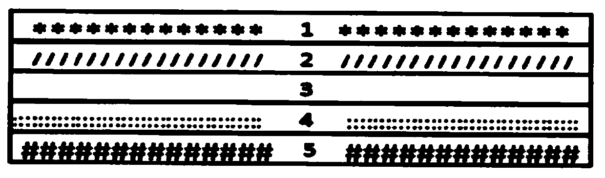
[0026] Such as figure 1 As shown, the pepsinogen I / pepsinogen II detection kit is a detection kit for fluorescence quantitative immunochromatography, including a detection card, and the detection card is sequentially provided with: PVC plate 1, sample pad 2, Combination pad 3, chromatographic membrane 4 and absorbent paper 5; wherein the chromatographic membrane includes a detection area and a quality control area; the detection area is coated with a detection area of ​​anti-PG I monoclonal antibody and PG II monoclonal antibody, for capturing antigen; The quality control area is coated with goat anti-mouse IgG antibody for signal detection. Above-mentioned chromatographic membrane 4 is nitrocellulose membrane, makes through the following steps:

[0027] 1) Using a cell line different from the anti-PG I and anti-PG II monoclonal antibody cell lines used on the binding pad 3, prepare and purify anti-human cardiac troponin I antibody and anti-myoglobin antibody by standard asci...

Embodiment 2

[0079] In order to improve the stability and sensitivity of the kit, the optimization measures taken also include:

[0080] The rare earth fluorescent microsphere-labeled anti-PG I monoclonal antibody and PG II monoclonal antibody-microsphere coupling complex adsorbed on the binding pad 3 were prepared by the following steps:

[0081] The aldylation step of rare earth fluorescent microspheres is as follows: take 5 mg of rare earth fluorescent microspheres, wash with 20 mM carbonate buffer solution with pH 9.5 for 3 times by centrifugation at 12000 rpm for 5 minutes, and finally resuspend Add 500ul of aldylated dextran and 0.2μg of 2-aminoethanesulfonic acid to 100ul of the above-mentioned carbonate buffer solution containing 1.5ul of thioglycolic acid monoethanolamine, mix well, and react in the dark at room temperature for 4 hours. Wash by centrifugation and resuspend in 100ul of the above-mentioned carbonate buffer, and place at 4°C for later use. The presence of monoethano...

Embodiment 3

[0083] Stability test

[0084] Stability of the test card: The test card in the kit is usually stored in a dry and cool environment at room temperature. The test card is placed in an oven at 50°C for one month to conduct an accelerated damage stability test, which is equivalent to the validity of the test card stored at room temperature for one year. Place the test card in an oven at 50°C for 1 week, 2 weeks, 3 weeks, and 4 weeks, then take it out and put it in a drying room, and compare it with the reagent card placed in the drying room at room temperature. card stability. The detection card of Example 2 was placed in an oven at 50°C for accelerated destruction for 1 week, 2 weeks, 3 weeks, and 4 weeks. The reaction curve in the case of the standard product test was basically consistent with the detection card stored at room temperature, and the fluorescence signal of low, medium and high concentrations The T / C value basically did not change, indicating that the fluorescenc...


PUM

PropertyMeasurementUnit
diameteraaaaaaaaaa

Abstract

The invention provides a pepsinogen I/pepsinogen II detection kit, and belongs to the technical field of fluorescence immunochromatography in medical immunology. The pepsinogen I/pepsinogen II detection kitwhich is a detection kit of fluorescence quantitative immunochromatography, comprises a detection card, and is characterized in that the detection card is provided with a PVC board, a sample pad, a combination pad, a chromatography membrane and a water absorption paper in sequence; and the chromatographic membrane comprises a detection region and a quality control region, a detection regionof an anti-PG I monoclonal antibody and a PG II monoclonal antibody are coated with the detection region, and a sheep anti-mouse IgG antibody is coated with the quality control region. Test samples ofthe reagent kit are human serum, plasma or whole blood samples, and good detection specificity, higher sensitivity, simple and convenient operation and stable fluorescent markers are achieved to ensure the accuracy of the detection.

Description

technical field [0001] The invention belongs to the technical field of fluorescence immunochromatography in medical immunology, and in particular relates to a pepsinogen I / pepsinogen II detection kit. Background technique [0002] Pepsinogen (PG) is a digestive enzyme precursor secreted by the stomach and an inactive precursor of pepsin in gastric juice. According to its distribution and immunogenicity, it can be divided into two subtypes, PG I and PG II. . PG I and PG II are good diagnostic indicators for Helicobacter pylori (HP) infection and atrophic gastritis, and their levels can reflect the secretory function of gastric mucosa. PG I mainly reflects the mucosal state of the gastric fundus and gastric body. Increased gastric acid secretion, gastritis and peptic ulcer PG I increase, decreased gastric acid secretion or gastric mucosal gland atrophy; when atrophic gastritis occurs, it will cause The principal cells of the gastric mucosa are lost, the secretory ability of ...

Claims


Application Information

Patent Timeline
Patent Type & Authority Applications(China)
IPC IPC(8): G01N33/533G01N33/573
CPCG01N33/533G01N33/573
Inventor 邓川郭亚楠张守涛陶新博杨永芳
Owner ZHEJIANG JUKANG BIOENG CO LTD
Why Eureka
Try Eureka