
Brain blood vessel endothelial cell line for quickly detecting activity of classical Wnt signal channel

A technology for cerebrovascular endothelial and endothelial cells, which is applied in the field of drug screening for rapid detection of the classic Wnt signaling pathway activity, can solve the problems of technical difficulty, time-consuming, and poor repeatability of the detection method, and achieve improved screening efficiency and accuracy, The effect of simple preparation method

Pending Publication Date: 2019-09-10
SHENZHEN INST OF ADVANCED TECH
View PDF5 Cites 3 Cited by
  • Summary
  • Abstract
  • Description
  • Claims
  • Application Information

AI Technical Summary

Problems solved by technology

[0014] The existing in vivo and in vitro experimental systems for detecting the activity of the canonical Wnt signaling pathway have significant limitations: 1) The in vivo mouse system is costly, time-consuming, and the detection method is technically difficult and poorly reproducible, requiring immunofluorescence staining or Immunohistochemistry is prone to false negatives or false positives, and cannot achieve high-throughput screening of a large number of candidate drug libraries; 2) The in vitro cell system can avoid the shortcomings of the above-mentioned in vivo mouse system, with low cost, high sensitivity, and easy operation. Convenient and high-throughput screening, however, the existing in vitro cell screening systems are non-cerebrovascular endothelial cells, and drugs screened based on these cell systems cannot reflect their effects on cerebrovascular endothelial cells
During the preparation process, although there are transfection experiments of HEK293 cell lines in the prior art, the existing HEK 293 cell lines and mouse L cell lines are easy to transfect, and it is easy to introduce exogenous DNA plasmids, while cerebrovascular endothelial cells Difficult to use common transfection reagents for transfection, must use lentiviral means, high technical requirements
At the same time, cerebrovascular endothelial cells are resistant to various antibiotics, and it is difficult to select the type or concentration of antibiotics

Method used


Image

  • Brain blood vessel endothelial cell line for quickly detecting activity of classical Wnt signal channel
  • Brain blood vessel endothelial cell line for quickly detecting activity of classical Wnt signal channel
  • Brain blood vessel endothelial cell line for quickly detecting activity of classical Wnt signal channel

Examples


Embodiment 1

[0069] Embodiment 1 preparatory work and pre-experiment

[0070] The bEnd.3 cell line was purchased from the American Type Culture Collection (ATCC). Cignal Lenti CMV Renilla Control (Hygro) and Cignal Lenti TCF / LEF Reporter (Luciferase; Puro) lentiviruses were purchased from Qiagen. Hygromycin and Puromycin were purchased from Sigma. Buy Dual from Promega- Reporter Assay System, used to detect the expression level of luciferase. Expand the culture of bEnd.3 cells, and freeze some bEnd.3 cells for preservation. Different concentrations of hygromycin ( figure 1 ) to treat the bEnd.3 cells for one week, and observe the morphology of the bEnd.3 cells under an ordinary optical microscope.

[0071] 100,000 bEnd.3 cells were evenly seeded into six-well plates, cultured overnight and the growth status of the cells was observed. Hygromycin was added when the cell density reached 80%. The concentrations of hygromycin were 0 μg / ml, 50 μg / ml, 100 μg / ml and 200 μg / ml, respectively...

Embodiment 2

[0073] Example 2 Construction of bEnd.3 cell line stably expressing Renilla

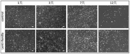
[0074] The promoter of Cignal Lenti CMV Renilla Control (Hygro) is CMV with hygromycin resistance. The bEnd.3 cells were infected with Lenti CMV Renilla virus for 72 hours. Then 100 μg / ml hygromycin was added to the cell culture medium for continuous selection for 12 days, and finally the cell line stably expressing Renilla was screened out ( image 3 ). The bEnd.3 cell line stably expressing Renilla was used as an internal reference fluorescence in the dual luciferase reporter gene experiment. The bEnd.3 cell line Renilla bEnd.3 cell line stably expressing Renilla was continued to be cultured in a medium containing hygromycin (50 μg / ml), the cultured cells were expanded, and a part of the cells were frozen for preservation.

Embodiment 3

[0075] Example 3 Construction of bEnd.3 cell line stably expressing Renilla / TOP flash

[0076] Cignal Lenti TCF / LEF Reporter (Puro) virus was added to the Renilla bEnd.3 cell line, and the cells were continuously infected for 72 hours. Lenti TCF / LEF Reporter (Puro) virus expresses TOP-Flash luciferase using the TCF / LEF promoter with puromycin resistance. Then add puromycin (4 μg / ml) and hygromycin (50 μg / ml) to the cell culture medium to select Renilla bEnd.3 stably transfected cell line for 3 days. Then reduce the concentration of antibiotics, add puromycin (1 μg / ml) and hygromycin (50 μg / ml) to the cell culture medium to select Renilla bEnd.3 cells for 10 days, and select a cell line stably expressing TOP-Flash / Renilla ( Figure 4 ). Continue to expand and cultivate stable cell lines, and freeze some cells for preservation.

[0077] 5.2 Effect examples

[0078] The bEnd.3 cell line stably transfected with TOP-Flash / Renilla was verified by using Wnt3a to activate the can...


PUM


Abstract

The invention provides a preparing method of a brain blood vessel endothelial cell line for stably expressing TOP-Flash / Renilla dual-luciferase reporter genes. The preparing method comprises the following steps of 1, selecting cerebral microvascular endothelial cells, infecting the cerebral microvascular endothelial cells with a slow virus carrying a Renilla gene, and conducting screening to obtain a cell line for stably expressing Renilla; 2, infecting the cell line obtained in step 1 with a slow virus carrying a TOP-Flash gene, and conducting screening to obtain the brain blood vessel endothelial cell line for simultaneously stably expressing the Renilla and TOP-Flash dual-luciferase reporter genes. The brain blood vessel endothelial cell line can be used for screening active substancesfor activating the activity of a Wnt signal channel in the brain blood vessel endothelial cells, the screening efficiency and accuracy are improved, and the preparing method is simple.

Description

technical field [0001] The invention relates to the field of biotechnology, in particular to a drug screening method for rapidly detecting the activity of the canonical Wnt signaling pathway. Background technique [0002] 1. Current status of stroke [0003] Cerebral stroke, also known as cerebral apoplexy, is an acute cerebrovascular disease caused by the sudden rupture or blockage of blood vessels in the brain, which leads to local cerebral blood supply disturbance and hypoxic-ischemic lesion necrosis of brain tissue, of which more than 75% are ischemic cerebral vascular diseases. stroke (1). Stroke has the characteristics of high incidence rate, high death and disability rate and high recurrence rate. Studies have shown that stroke is the leading cause of death and disability among residents in my country, with about 2.7 million new patients each year, and the trend is increasing year by year (2,3). Stroke brings a heavy economic burden to the whole society and patient...

Claims


Application Information

Patent Timeline
Patent Type & Authority Applications(China)
IPC IPC(8): C12N15/867C12N5/10C12Q1/02
CPCC12N15/86C12N5/069G01N33/5041G01N33/5064C12N2740/15043C12N2800/107C12N2510/00C12N2503/02G01N2500/10
Inventor 畅君雷王田喜尹美芳丘林辉
Owner SHENZHEN INST OF ADVANCED TECH
Learn More