
Application of tuberculosis immunohistochemical kit in diagnosis of tuberculosis pathological tissues

A technology of immunohistochemistry and reagent kits, which is applied in the field of tuberculosis diagnosis, can solve the problems of cumbersome false positive rate, high cost, cumbersome operation, etc., to improve the intensity and range of color signal, improve the diagnosis rate and distribution space wide effect

Inactive Publication Date: 2019-11-26
武汉顺可达生物科技有限公司
View PDF5 Cites 0 Cited by
  • Summary
  • Abstract
  • Description
  • Claims
  • Application Information

AI Technical Summary

Problems solved by technology

At present, the diagnostic techniques for tuberculosis clinically used mainly include tuberculin (PPD) test, acid-fast staining, serological antibody detection, ELISpot detection of IFN-γ, and gene chips. There are more or less limitations in clinical application. Limitations, such as high false positive rate, cumbersome operation, etc., especially these methods cannot be used as the basis for the diagnosis of tuberculosis; and the sputum bacterial culture and PCR methods that can be used as the basis for the diagnosis of tuberculosis: culture is also cumbersome and time-consuming, so It is often unable to guide clinical treatment. In addition, due to various reasons, the positive rate of tuberculosis culture in my country is currently low, which affects the timely and accurate diagnosis of tuberculosis. The PCR method is relatively time-consuming and short, but there is a high cost, and it cannot distinguish between past infections and new infections. Tuberculosis
No immunohistochemical method targeting tuberculosis surface glycolipids has been reported

Method used


Image

  • Application of tuberculosis immunohistochemical kit in diagnosis of tuberculosis pathological tissues
  • Application of tuberculosis immunohistochemical kit in diagnosis of tuberculosis pathological tissues
  • Application of tuberculosis immunohistochemical kit in diagnosis of tuberculosis pathological tissues

Examples


Embodiment 1

[0023] Embodiment 1 kit composition

[0024] 1. PBS formula (concentration is 0.01M, pH value is 7.4): 2.9g Na 2 HPO 4 12H 2 O, 0.3g NaH 2 PO 4 2H 2 O and 9g NaCl were dissolved in 1L of distilled water to adjust the pH value to 7.4.

[0025] 2. PBST formula: on the basis of the above PBS, add Tween-20 with a final concentration of 0.025% and mix well.

[0026] 3. Sodium citrate buffer (10 mM sodium citrate, 0.05% Tween-20, pH 6.0).

[0027] 4. Aptamer "antibody" (300nM): screened by SELEX, its nucleotide sequence is shown in SEQ ID No.1, with ddH 2 O adjusted the final concentration to 300 nM.

[0028] 5. Streptavidin-HRP: Diluted at 1:500 for use.

[0029] 6. DAB staining solution: first prepare 20×DAB dyeing mother solution: pour 0.1g of diaminobenzidine (3,3'-diaminobenzidine, DAB) into 10mL of distilled water to obtain a mixed solution, add 3-5 drops to the mixed solution 10M HCl until the mixture turns light brown, then the DAB is completely dissolved, aliquote...

Embodiment 2

[0033] The making of embodiment 2 tissue sections (taking paraffin embedding as example)

[0034] All sliced ​​tissues were obtained from patients who were hospitalized for surgery from January 2018 to August 2018 at Wuhan Jinyintan Hospital (Wuhan Medical Treatment Center).

[0035] All specimens were confirmed by pathology, and sliced ​​specimens were taken from the affected parts. All specimens were obtained from the Pathology Department of Wuhan Jinyintan Hospital (Wuhan Medical Treatment Center). All tissue samples were routinely fixed in 10% neutral formalin, embedded in paraffin, and the paraffin block was screened without obvious defects, and stored at room temperature for future use.

[0036] The main process of making tissue slices is as follows:

[0037] (1) Tissue (thickness<3mm) blocks were fixed overnight with 10% neutral formalin.

[0038] (2) Rinse the slices with tap water for 5 minutes to remove excess formalin.

[0039] (3) Immerse the slices in 70% ethan...

Embodiment 3

[0045]Example 3 Immunohistochemical staining + acid-fast staining + hematoxylin counterstaining:

[0046] (1) Paraffin sections were dewaxed and rehydrated: put the paraffin sections in a 60-degree oven for 30 minutes. Xylene I and II were dewaxed for 5 minutes, put into 10%, 95%, 90%, 80%, and 70% methanol for 2 minutes each, and then put into distilled water for 5 minutes.

[0047] (2) Add 100 μL 3% H 2 o 2 Incubate at room temperature for 5-10 minutes to eliminate endogenous peroxidase activity.

[0048] (3) Antigen retrieval: Add sodium citrate buffer solution (10 mM sodium citrate, 0.05% Tween-20, pH 6.0) into a beaker and heat to boiling in a water bath. Put the slices into the slide rack and put them in a beaker to boil the slices for 30 minutes. Take the beaker out of the water bath and let it cool down to room temperature naturally.

[0049] (4) PBS (1 mL) washed the sections twice.

[0050] (5) Block with 10% normal goat serum (diluted in 0.5% PBS), and incubat...


PUM


Abstract

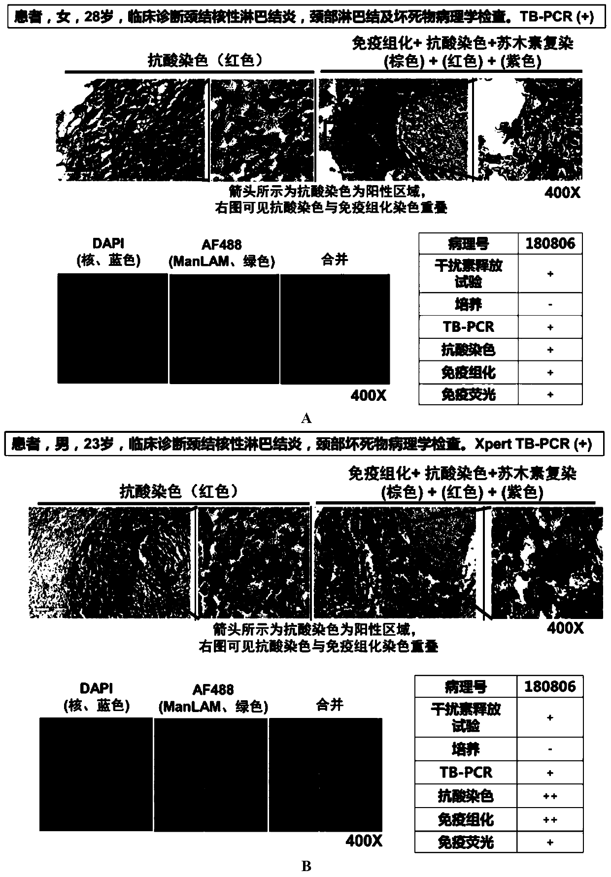
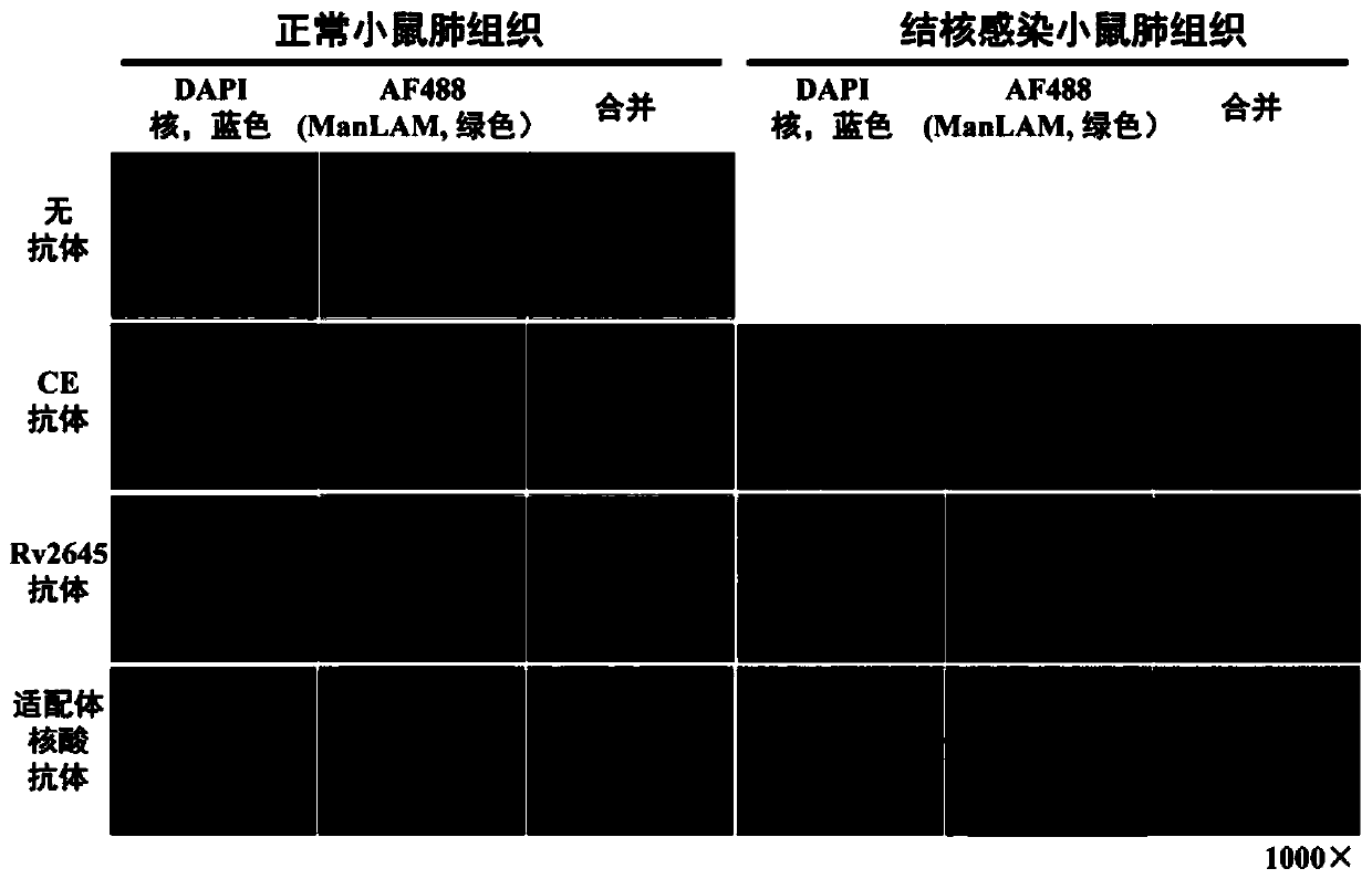
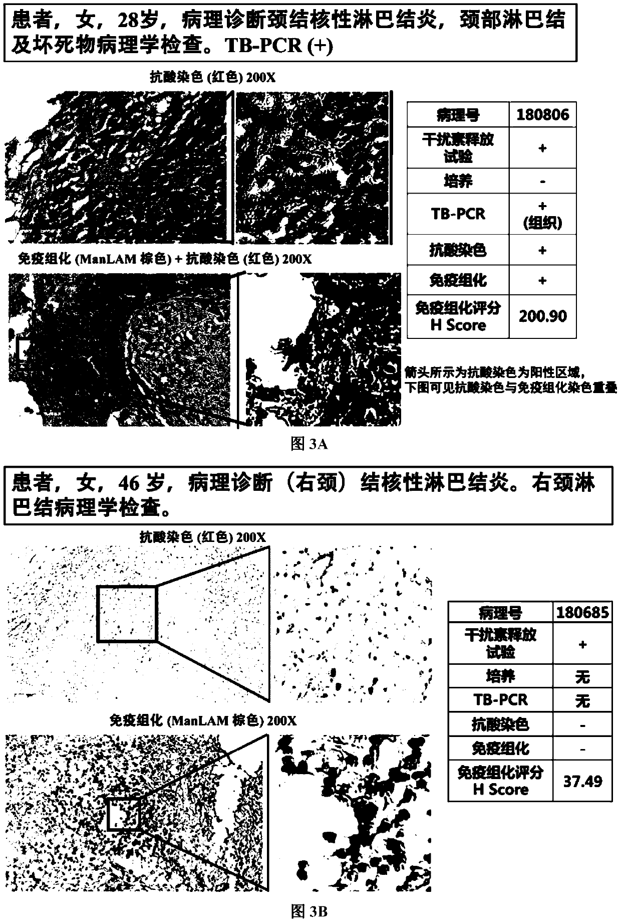
The invention discloses an application of a tuberculosis immunohistochemical kit in diagnosis of tuberculosis pathological tissue (including intrapulmonary and extrapulmonary tuberculosis pathologicaltissues), and belongs to the field of tuberculosis diagnosis. According to the invention, a tuberculosis antigen specific aptamer nucleic acid antibody is adopted, biotin, or fluorescence, chemiluminiscence and the like are adopted for labeling, mycobacterium tuberculosis specific antigen (ManLAM antigen) existing in tuberculosis pathological tissues is detected, and the antigen can exist in thalli and be secreted to the surrounding space; clinical sample immunohistochemistry proves that the tuberculosis antigen specific aptamer antibody is high in staining on tuberculosis infected tissues and low in background staining. The tuberculosis is tuberculosis caused by mycobacterium tuberculosis or drug-resistant mycobacterium tuberculosis (including intrapulmonary tuberculosis and extrapulmonary tuberculosis) and all tuberculosis co-affected by people and livestock. The invention provides a tuberculosis immunohistochemical kit in tuberculosis pathological tissues, and provides a new tool for histopathological definite diagnosis of tuberculosis pathological tissues.

Description

technical field [0001] The present invention relates to the field of tuberculosis diagnosis, and provides the application of a tuberculosis immunohistochemical kit in the diagnosis of tuberculosis (including intrapulmonary and extrapulmonary tuberculosis) lesion tissue, which provides the histopathological diagnosis of tuberculosis and zoonotic tuberculosis lesion tissue new tools. Background technique [0002] Tuberculosis is still a chronic infectious disease that seriously endangers human health, and tuberculosis caused by Mycobacterium tuberculosis is a zoonotic tuberculosis. Nearly one-third of the world's population is currently infected with Mycobacterium tuberculosis. According to WHO's "Global Tuberculosis Report 2018," it is estimated that there will be 10 million new tuberculosis cases in 2017, and 1.3 million deaths due to tuberculosis, exceeding the death toll from other infectious diseases. sum. [0003] Early and clear diagnosis is extremely important for tu...

Claims


Application Information

Patent Timeline
IPC IPC(8): G01N33/533G01N33/541G01N33/569G01N1/30G01N1/28
CPCG01N33/533G01N33/541G01N33/569G01N1/30G01N1/286G01N2001/2873
Inventor 章晓联
Owner 武汉顺可达生物科技有限公司
Learn More