3D bioprinting ink, preparation method of ink, tissue engineering scaffold and preparation method of scaffold
A tissue engineering scaffold and bioprinting technology, applied in the field of medicine, can solve the problems of increasing the cost of preparation and increasing the complexity of the production process, and achieve the effects of reducing treatment costs, relieving patients' pain, and improving strength
- Summary
- Abstract
- Description
- Claims
- Application Information
AI Technical Summary
Problems solved by technology
Method used
Image

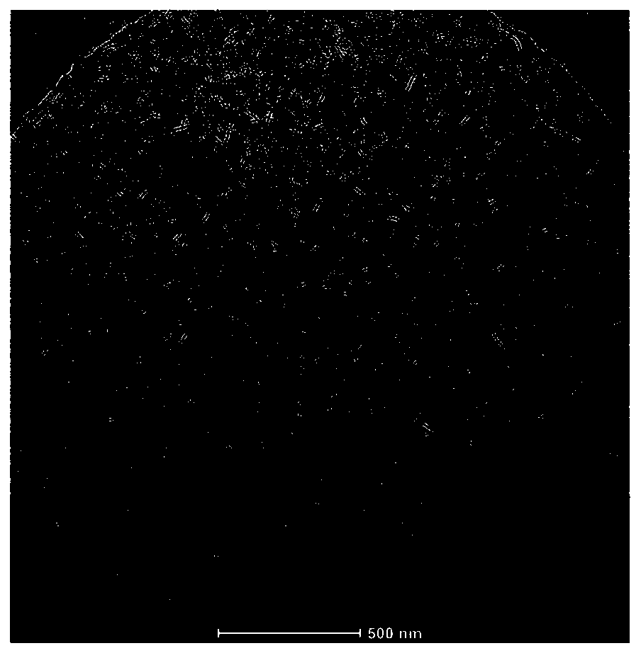
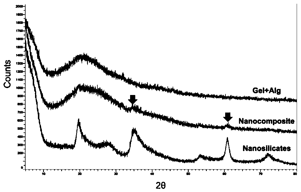
Examples
preparation example Construction
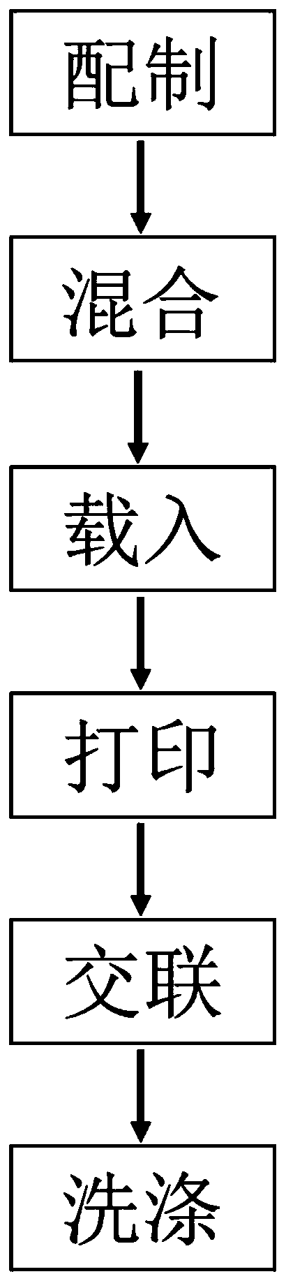
[0042] A preparation method for 3D bioprinting ink, comprising the following steps:
[0043] S1, weighing raw materials
[0044] Weigh the gelatin particles, sodium alginate powder and lithium magnesium silicate powder according to the set amount, and sterilize and disinfect the gelatin particles, sodium alginate powder and lithium magnesium silicate powder;
[0045] S2, preparation of raw materials
[0046] Dissolve the gelatin particles obtained in S1 with sterile deionized water to obtain a gelatin solution, add sodium alginate powder to the gelatin solution, and obtain a mixed prepolymer solution of gelatin-sodium alginate after dissolving, dissolve the lithium magnesium silicate powder in In sterile deionized water, lithium magnesium silicate colloid was obtained;
[0047] S3, mixed
[0048] Mix the gelatin-sodium alginate mixed prepolymer solution obtained in S2 with the lithium magnesium silicate colloid in equal volumes to obtain a nanocomposite hydrogel, and combin...
Embodiment 1
[0060] The formulations of bioactive tissue engineering bone scaffolds listed in this example include nanocomposite hydrogels and human bone marrow mesenchymal stem cells, wherein the nanocomposite hydrogels include gelatin, sodium alginate, nanoscale lithium magnesium silicate and Deionized water; nanocomposite hydrogel including gelatin 70mg / mL~100mg / mL, sodium alginate 10mg / mL~30mg / mL, magnesium silicate lithium 10mg / mL~30mg / mL, among them, human bone marrow mesenchymal stem cells The volume ratio of the suspension to the nanocomposite hydrogel was 1:400.
[0061] Such as figure 1 Shown, as a preferred embodiment of the present invention, specifically carry out according to the following steps:
[0062] Step 1. Preparation of raw materials: First, put the pre-weighed gelatin granules, sodium alginate powder and lithium magnesium silicate powder in a sterile centrifuge tube, sterilize with ethylene oxide, and set aside; add the gelatin granules to the Put a corresponding v...
PUM
| Property | Measurement | Unit |
|---|---|---|
| diameter | aaaaa | aaaaa |
| thickness | aaaaa | aaaaa |
| concentration | aaaaa | aaaaa |
Abstract
Description
Claims
Application Information