Small-caliber vascular stent for decellularization and preparation method of small-caliber vascular stent

A vascular stent and small-caliber technology, which is applied in the field of preparation of decellularized small-caliber vascular stents, can solve the problems of inflammatory immune rejection, changes in the mechanical properties of the decellularized solution, etc., to reduce immunogenicity, reduce nuclear substances and antigenic components, Conducive to the effect of adhesion growth, proliferation and migration

Pending Publication Date: 2020-01-31
GUANGDONG GENERAL HOSPITAL
View PDF12 Cites 0 Cited by
  • Summary
  • Abstract
  • Description
  • Claims
  • Application Information

AI Technical Summary

Problems solved by technology

[0005] The present invention provides a method for preparing decellularized small-caliber vascular stents to solve the problems of inflammatory immune rejection, changes in mechanical properties by decellularization schemes, and recellularization

Method used


Image

  • Small-caliber vascular stent for decellularization and preparation method of small-caliber vascular stent
  • Small-caliber vascular stent for decellularization and preparation method of small-caliber vascular stent
  • Small-caliber vascular stent for decellularization and preparation method of small-caliber vascular stent

Examples


Embodiment 1

[0036] Example 1 Preparation method of decellularized human umbilical artery stent

[0037] Experimental material: human umbilical artery

[0038] Equipment: ultra-clean table, shaker, sterile biochemical incubator

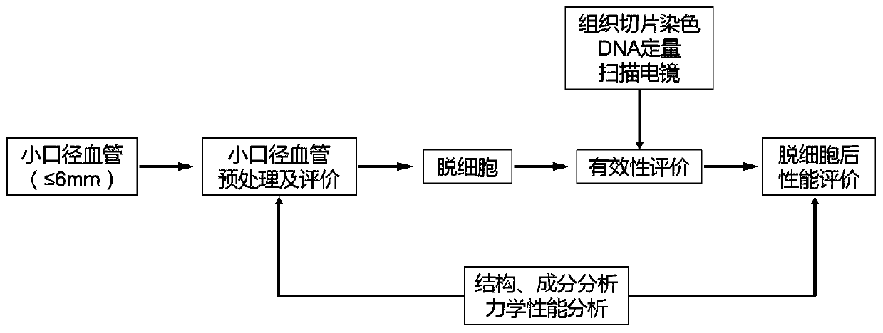
[0039] The specific cultivation method includes the following steps:

[0040](1) Obtaining of umbilical artery: The umbilical cords of newborns with good health and gestational age (38-42 weeks) were obtained, and the umbilical cords were obtained with the informed consent of the puerpera and voluntary donation (the research protocol was approved by the Ethics Committee of Guangdong Provincial People’s Hospital). 4-6cm, diameter 2-3mm, the following operations are all performed under the sterile environment of ultra-clean bench and biochemical incubator;

[0041] (2) Pretreatment of the umbilical artery: carefully peel off the connective tissue around the umbilical artery to avoid damage to the adventitia of the umbilical artery, rinse the separated umbilical ar...

Embodiment 2

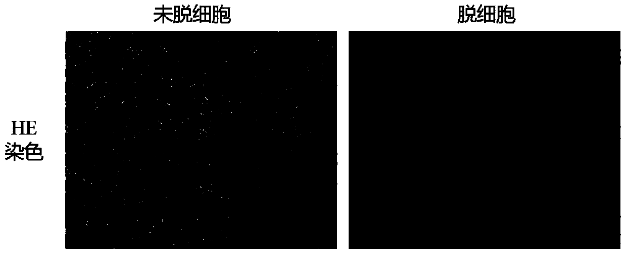
[0045] Example 2 Evaluation of the Effectiveness of the Decellularized Human Umbilical Artery Stent

[0046] HE staining of human umbilical arteries before and after decellularization

[0047] The decellularized umbilical artery stent prepared in the embodiment of the present invention and the umbilical artery of the control group not treated with chemical detergent were subjected to HE staining, and the specific steps were as follows: two groups of umbilical arteries were immersed in 4% paraformaldehyde and fixed overnight, The tissue was taken out the next day, washed twice with PBS, 15min / time, dehydrated with 50%, 70%, 80%, 90%, 95%, 100%, 100% ethanol gradient, then immersed in xylene for 20min; The umbilical arteries of the group were fully immersed in hot-melted paraffin (54°C-56°C), embedded in paraffin in the mold, and after the paraffin solidified after cooling, the embedding block was taken out, paraffin sectioned, cut into 4mm, and exhibited in a water bath at 40°C...

Embodiment 3

[0050] Example 3 Component and Structure Detection of Decellularized Human Umbilical Artery Stent

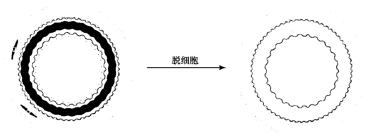
[0051] Scanning electron microscopy of human umbilical arteries before and after decellularization

[0052] The decellularized umbilical artery stent prepared in the embodiment of the present invention and the umbilical artery of the control group not treated with chemical detergent were subjected to scanning electron microscope experiments. The specific steps were as follows: two groups of umbilical arteries were placed in 2.5% glutaraldehyde solution Fix at 4°C for 4h, discard the fixative, rinse the sample with PBS, then dehydrate with ethanol gradient, replace the ethanol twice with isoamyl acetate, freeze the specimen at -20°C, -40°C, and -80°C for 12h, and place them in a freeze dryer. Dry in medium for 12h, and prepare the specimens for later use. Metal spraying was carried out before observation with a scanning electron microscope. In order to make the sample conductive...


PUM

PropertyMeasurementUnit
diameteraaaaaaaaaa

Abstract

The invention discloses a small-caliber vascular stent for decellularization. The small-caliber vascular stent is characterized in that antigenic components such as cells and nuclear matter on blood vessels are fully removed, the immunological rejection of blood vessels is reduced to a greater extent, and the blood vessels have good compliance; extracellular matrix components of small-caliber blood vessels are better retained; conformation and space structure destruction of original structural protein such as collagen fiber and elastic fiber of blood vessel tissue are maintained, so that bloodvessel tissue has mechanical properties and mechanical strength similar to that before decellularization; proper fiber pores of micro pore materials formed after decellularization provide natural microenvironments, adherent growth, proliferation and migration of host cells are facilitated, and cytotoxicity is avoided; and the materials are modified after decellularization to reduce thrombosis andincrease forward patency rate of the small-caliber vascular stent. The obtained small-caliber vascular stent for decellularization has good repair, growth and reconstruction functions.

Description

technical field [0001] The invention belongs to the field of tissue engineering tubular materials, and in particular relates to a method for preparing a decellularized small-caliber vascular stent. Background technique [0002] Vascular disease, especially atherosclerosis, is one of the leading causes of mortality and morbidity in Western societies and is therefore made of synthetic polymers including a variety of artificial materials including Dacron and expanded polytetrafluoroethylene (ePTFE) Vascular grafts based on these materials have been used in the transplantation of large and medium-caliber blood vessels (diameter>6mm), but the high thrombosis rate and low long-term patency rate after transplantation limit these materials to be used as small-caliber artificial blood vessels. [0003] Autologous blood vessels are currently the best choice for vascular bypass graft materials. However, limited by the number of available autologous blood vessels and the progression ...

Claims


Application Information

Patent Timeline
Patent Type & Authority Applications(China)
IPC IPC(8): A61L27/36A61L27/50
CPCA61L27/507A61L27/3625A61L27/3687A61L2430/40A61L2400/18
Inventor 林展翼梁远锋刘尚敏李东风姚博谦
Owner GUANGDONG GENERAL HOSPITAL
Why Eureka
Try Eureka