
Improved microcarrier living cell fluorescence staining method

A fluorescent staining and microcarrier technology, applied in the biological field, can solve the problems of low cell viability, cell shedding, low cell permeability, etc.

Inactive Publication Date: 2020-05-19
浙江卫未生物医药科技有限公司
View PDF7 Cites 3 Cited by
  • Summary
  • Abstract
  • Description
  • Claims
  • Application Information

AI Technical Summary

Problems solved by technology

[0004] The above staining methods utilize the difference in cell membrane permeability of living and dead cells and the difference in the binding ability of dyes and nucleic acid substances or the difference in metabolism of living and dead cells to stain living and dead cells respectively. However, these staining methods have three common shortcomings, namely One is that the cell permeability is low, and it is difficult for the dye to fully combine with the substances in the cell to emit strong fluorescence, resulting in a low cell viability; the other is that the difference between the osmotic pressure of the dye and the pH of the cell growth environment easily leads to the cell from the microcarrier. The surface falls off; the third is that the fluorescence emitted by the dye combined with the cells is easily quenched, resulting in inaccurate live cell counts

Method used


Image

  • Improved microcarrier living cell fluorescence staining method
  • Improved microcarrier living cell fluorescence staining method
  • Improved microcarrier living cell fluorescence staining method

Examples


Embodiment 1

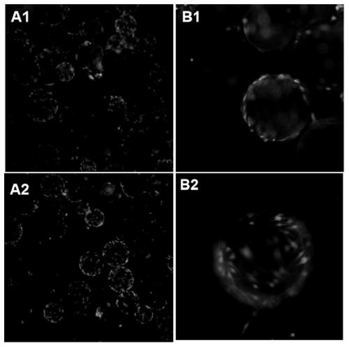
[0019] Human adipose-derived mesenchymal stem cells (ADSCs) were cultured with Cephodex spherical microcarriers (Binzhou Bell Carey Biotechnology Co., Ltd., Shandong), and the microcarriers with a concentration of 3 g / L were pre-expanded, sterilized, and sterilized according to the instructions. processing, the specific method is as follows:

[0020] Take 0.3g of microcarriers and add 30-50ml of sterile PBS to soak for 48 hours to fully expand, sterilize by high-pressure steam for 30 minutes, wash with sterile PBS for 3 times, add 50ml of complete medium into a specific bioreactor, and incubate at 37°C for 24 hours , for sterility testing. After confirming the sterility, discard the test solution, add 100ml of complete medium again, and divide the ADSCs according to the cell density of 3*10 5 / ml for inoculation and cultured with intermittent stirring.

[0021] When the cells adhere to the wall to about 80%, take 1ml of microcarrier cell suspension and put them into 24-well ...

Embodiment 2

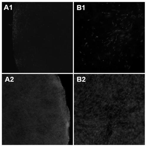
[0023] Human umbilical cord mesenchymal stem cells (hU-MSCs) were cultured using fixed-bed Cephodisk sheet microcarriers (Binzhou Bell Carey Biotechnology Co., Ltd., Shandong). The specific culture method was: take 100 microcarriers and add 30-50ml sterile Soak in DPBS for 30 minutes, sterilize with high-pressure steam for 30 minutes, wash with sterile DPBS for 3 times, add 20ml of complete medium into a specific culture bottle, incubate at 37°C for 24 hours, perform sterility test, and discard the test after confirming sterility solution, re-add 20ml of complete medium, hU-MSCs according to cell density 1.5-2.5*10 4 / ml for inoculation and cultured with low-speed intermittent agitation. On the third day of culture, set up group A and group B, and randomly select one piece of microcarriers for living cell fluorescence staining observation, the specific method is as follows:

[0024] Group A washed the microcarriers with sterile DPBS for 1-2 times, added 2ml of Calcein-AM stai...


PUM


Abstract

The invention relates to the technical field of biology, in particular to a living cell fluorescence staining method for culturing cells by using a microcarrier technology. The method is used for identifying the activity of cells on a microcarrier. Before staining, a cell membrane permeable agent triton X-100 is added to enhance cell permeability and promote dye to enter cells to be combined withnucleic acid; during the staining, a serum-free culture medium is used as a buffer solution to prepare a calcein-methyl acetate (Ceclein-AM) living cell staining solution which is closer to a cell growth environment and prevents cells from falling off; after the staining, an anti-fluorescence attenuation agent DABCO, namely 1, 4-diazobicyclo[2, 2, 2]-octane, is added, and the fluorescence emittingtime is effectively prolonged such that the detection result is more accurate. By using the method, the activity of adherent cells on the microcarrier can be judged according to the intensity of green fluorescence, and meanwhile, the growth of the cells is not influenced.

Description

technical field [0001] The invention relates to the field of biotechnology, in particular to a method for fluorescent staining of living cells cultured on microcarriers. Background technique [0002] Microcarriers are generally composed of natural dextran or various synthetic polymers, which are suitable for the growth of adherent cells. The basic types mainly include liquid microcarriers, macroporous gelatin microcarriers, polystyrene microcarriers, and PHEMA microcarriers. Carriers, chitin microcarriers, polyurethane foam microcarriers, alginate gel microcarriers and magnetic microcarriers, etc. Commonly used commercial microcarriers mainly include Cytodex1, 2, 3, Cytopore and Cytoline. The basic principle of microcarrier cell culture technology is to add the harmless microcarrier to the culture medium of the culture container as a carrier to make the cells attach and grow on the surface of the microcarrier, and at the same time keep the microcarrier in a suspended state ...

Claims


Application Information

Patent Timeline
Patent Type & Authority Applications(China)
IPC IPC(8): G01N1/30
CPCG01N1/30
Inventor 陈锦阳李静静刘军权
Owner 浙江卫未生物医药科技有限公司
Learn More