
Preparation method of high-mutation urine stem cells in mitochondria mt.3243A > G mutation crowds
A technology of mt.3243a and mitochondria, applied in the field of biochemistry, can solve problems such as difficulties in establishing biological models
- Summary
- Abstract
- Description
- Claims
- Application Information
AI Technical Summary
Problems solved by technology
Method used
Image

Examples
Embodiment 1
[0034] Isolating and culturing stem cells from the urine of mutation patients and monoclonal isolation and culture, including the following steps:
[0035] 1) Add 5ml of a mixed solution of penicillin, streptomycin and amphotericin B (the content of penicillin is 10kU / ml, the content of streptomycin is 10mg / ml, and the content of amphotericin B is 25μg / ml) into the T75 bottle. Spray disinfectant around;
[0036] 2) Select the urine of the mitochondrial mt.3243A>G mutant population, and use the T75 bottle in step 1) for urine collection;
[0037] 3) Open the T75 bottle, add 50ml of urine sample to each of the four 50ml centrifuge red tubes, and centrifuge;
[0038] 4) Use a pipette to suck up the supernatant to less than 5ml in each red tube, add 15ml~20ml PBS to the first red tube, pipette and mix well, then transfer the resuspension to the next red tube, pipette again Mix well, and so on, until finally all the sediments of the tubes are collected in one tube, and centrifuge...
Embodiment 2
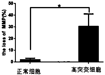
[0054] Example 2 Membrane Potential Detection
[0055] The amount of JC-1 staining working solution is 1ml, take an appropriate amount of JC-1 (200X), and dilute JC-1 by adding 8ml of ultrapure water to every 50μl of JC-1 (200X). Vigorously dissolve Vortex and mix JC-1 thoroughly. Then add 2ml of JC-1 staining buffer (5X), and mix well to get the JC-1 staining working solution.
[0056] a. For one well of the six-well plate, suck off the culture medium, wash the cells once with PBS or other appropriate solution if necessary according to the specific experiment, and add 1ml of mesenchymal stem cell culture medium. Cell culture medium may contain serum and phenol red.
[0057] b. Add 1ml JC-1 staining working solution and mix well. Incubate at 37°C for 20 minutes in a cell culture incubator.
[0058] c. During the incubation period, prepare an appropriate amount of JC-1 staining buffer (1X) by adding 4ml of distilled water to every 1ml of JC-1 staining buffer (5X), and place...
Embodiment 3
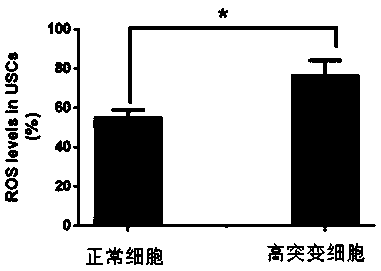
[0062] Example 3 ROS detection
[0063] Loading probes after collecting urine mesenchymal stem cells: Dilute DCFH-DA with serum-free medium according to 1:1000, so that the final concentration is 10umol / L.
[0064] Centrifuge the cells at 600g for 4min (or 3000rpm / 5min), remove the supernatant, suspend the cells in diluted DCFH-DA (1ml), the cell concentration is 10^6-2*10^7 / ml, and culture the cells at 37°C Incubate in the box for 20 minutes. Mix by inverting every 3-5 minutes to make the probe fully contact with the cells. The cells were centrifuged at 600 g for 4 min, and the cells were washed (centrifuged, broken, and blown) three times with serum-free cell culture medium to fully remove DCFH-DA that did not enter the cells. Add about 500ul (depending on the cell density) of culture medium, blow and mix well, smear 10ul sample onto the glass slide, and observe under the fluorescence microscope. Each sample has three fields of view, and each field of view is first shot wi...
PUM
 Abstract
Description
Claims
Application Information