Method for non-invasive preimplantation hereditary detection of embryos

A pre-implantation, genetic technology, applied in biochemical equipment and methods, microbial measurement/inspection, etc., can solve the problems of large volume range changes, low free DNA concentration, and complex culture medium components, etc., to achieve improvement The effects of data quality, good amplification uniformity, and avoiding sampling risks

Active Publication Date: 2020-07-24
阿吉安(福州)基因医学检验实验室有限公司
View PDF6 Cites 9 Cited by
  • Summary
  • Abstract
  • Description
  • Claims
  • Application Information

AI Technical Summary

Problems solved by technology

However, in the detection of maternal contamination, if the blastocyst culture medium is used for non-invasive detection, the concentration of free DNA in the blastocyst culture medium is extremely low, the volume range varies greatly (10-20 μL), and the composition of the culture medium is complex. Some substances may inhibit WGA amplification
Existing detection kits and detection methods cannot effectively avoid the above problems

Method used


Image

  • Method for non-invasive preimplantation hereditary detection of embryos
  • Method for non-invasive preimplantation hereditary detection of embryos
  • Method for non-invasive preimplantation hereditary detection of embryos

Examples


Embodiment 1

[0038] A kit for noninvasive preimplantation genetic testing consisting of the following reagents:

[0039] Pre-amplification mixture: 0.7mM dNTP, 40mM Tris-HCl (pH=7.5), 10mM MgCl 2 , 10% DMSO, 6uM primers, 1 μL high-fidelity DNA polymerase (1U / μL) (TAKARA);

[0040] Amplification mixture: 0.6mM dNTP, 40mM Tris-HCl (pH=7.5), 5mM MgCl 2 , 8% DMSO, 2.5uM primers, 5U high-fidelity DNA polymerase (1U / μL) (TAKARA);

[0041] Lysis solution: 25mM Tris-HCl (pH=8.3), 0.05% Triton X-100, 1.5mM EDTA;

[0042] Lyase: 2mg / mL proteinase K;

[0043] End repair reaction solution; end repair enzyme; DNA ligation buffer; DNA ligase; adapter; PCR amplification reaction solution; universal primers; index primers; deionized water.

experiment example 1

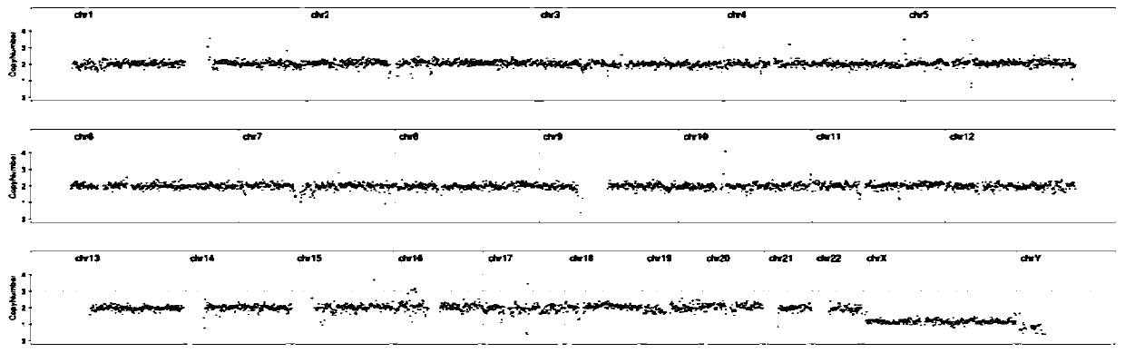
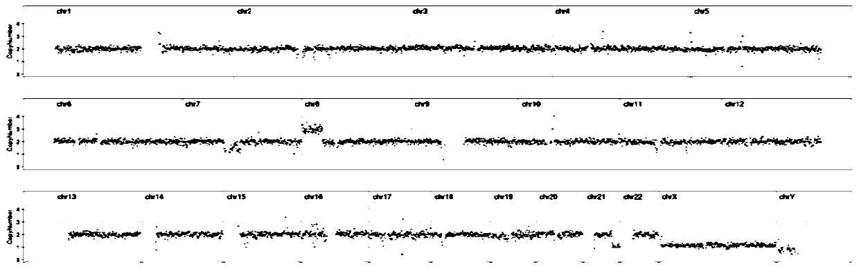
[0052] The two kits of Comparative Example 1 and Comparative Example 1 performed whole genome amplification, library preparation and on-machine sequencing on the same sample respectively. The amplification effects were compared, and the results are shown in Table 1. figure 1 and figure 2 shown.

[0053] Table 1

[0054] sample WGA concentration value (ng / ul) Comparative example 1 28.5 Example 1 46.7

[0055] Through the optimization of the kit in this application, it can be seen that the WGA concentration of the optimized kit in this application is significantly higher than that before optimization, and the CNV result dispersion of sequencing results after optimization is significantly better than that after optimization.

Embodiment 2

[0057] Embryo samples fertilized by intracytoplasmic sperm injection were cultured to the blastocyst stage, and conventional PGT-A blastocyst trophoblast cell biopsy and blastocyst culture fluid were used to detect chromosomal aneuploidy to evaluate whether the embryos were chromosomally normal.

[0058] Specific steps are as follows:

[0059] 1. Obtain samples of blastocyst culture fluid

[0060] After ICSI insemination, the fertilized eggs were cultured to the blastocyst stage, the blastocyst culture fluid was aspirated with a clean pipette, and 5-20 μL of the culture fluid was used as a sample for chromosomal aneuploidy detection. After aspirating the culture medium, the embryos were biopsied, and blastocyst trophoblast cells were taken for PGT-A detection.

[0061] 2. Whole genome amplification in blastocyst culture medium

[0062] 2.1 Lysis of blastocyst culture fluid samples

[0063] Add 10-20 μL of lysate and 0.6-1.2 μL of lyase to the culture solution sample tube co...


PUM


Abstract

The invention discloses a method for non-invasive preimplantation hereditary detection of embryos, and belongs to the technical field of biological detection. The method comprises the following steps:carrying out whole genome amplification on a blastocyst culture solution sample by using a kit, carrying out short tandem repeat sequence analysis on an amplification product and DNA samples of parents to detect maternal pollution, carrying out library preparation and next-generation sequencing detection on the amplification product to determine whether the number of chromosomes is abnormal or not; and optimizing a pre-amplification mixed solution and an amplification mixed solution by the provided kit. According to the method provided by the invention, the blastocyst culture solution can besubjected to parent source pollution detection, so that whether the chromosome aneuploidy detection result of the culture solution is accurate and reliable or not is judged. The invention provides a detection method for judging whether granular cells are completely removed or not, and the inhibition effect of components in the culture solution on amplification is effectively avoided through optimization of the kit, so that amplification uniformity is good, and the single cell amplification yield is high. The detection method is simple, the result is accurate, and data quality is improved.

Description

technical field [0001] The invention belongs to the technical field of biological detection, and in particular relates to a method for non-invasive preimplantation genetic detection of embryos. Background technique [0002] With the rapid development of human assisted reproductive technology, artificial insemination (AI), in vitro fertilization-embryo transfer (In Vitro Fertilization and Embryo Transfer, IVF-ET) and intracytoplasmic sperm injection (Intra-Cytoplasmic Sperm Injection, ICSI) has entered large-scale clinical application. About 50% of the embryos formed by in vitro fertilization have chromosomal abnormalities, which can lead to the loss of early embryos after transplantation, spontaneous abortion and stillbirth, which is one of the important reasons that limit the success rate and effective promotion of assisted reproductive technology. The traditional embryo screening method is to grade the quality of embryos through static observation of embryo morphology. Th...

Claims


Application Information

Patent Timeline
Patent Type & Authority Applications(China)
IPC IPC(8): C12Q1/6883C12Q1/6869
CPCC12Q1/6883C12Q1/6869C12Q2535/122
Inventor 郭文浒段佳丽陈浩
Owner 阿吉安(福州)基因医学检验实验室有限公司
Why Eureka
Try Eureka