GDF15 rapid quantitative fluorescent microsphere detection device and preparation method thereof

A technology of fluorescent microspheres and detection devices, which is used in measurement devices, biological tests, material inspection products, etc., can solve the problem of weak binding force, easy quenching of fluorescence, insufficient amount of protein adsorbed by NC membrane, and inability to detect and monitor GDF15 concentration. and other problems, to achieve the effect of improving sensitivity, increasing complexity, and novel structure

Active Publication Date: 2020-09-08
JILIN PROVINCE GERUISITE BIOTECHNOLOGY CO LTD
View PDF6 Cites 4 Cited by
  • Summary
  • Abstract
  • Description
  • Claims
  • Application Information

AI Technical Summary

Problems solved by technology

[0009] The present invention provides a GDF15 rapid quantitative fluorescent microsphere detection device and its preparation method to solve the problems that GDF15 concentration detection and monitoring cannot be realized without borrowing large instruments, fluorescence is easily quenched, and the amount of protein adsorbed by NC membrane is insufficient and the binding force is not strong question

Method used


Image

  • GDF15 rapid quantitative fluorescent microsphere detection device and preparation method thereof
  • GDF15 rapid quantitative fluorescent microsphere detection device and preparation method thereof
  • GDF15 rapid quantitative fluorescent microsphere detection device and preparation method thereof

Examples


Embodiment 1

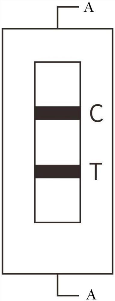
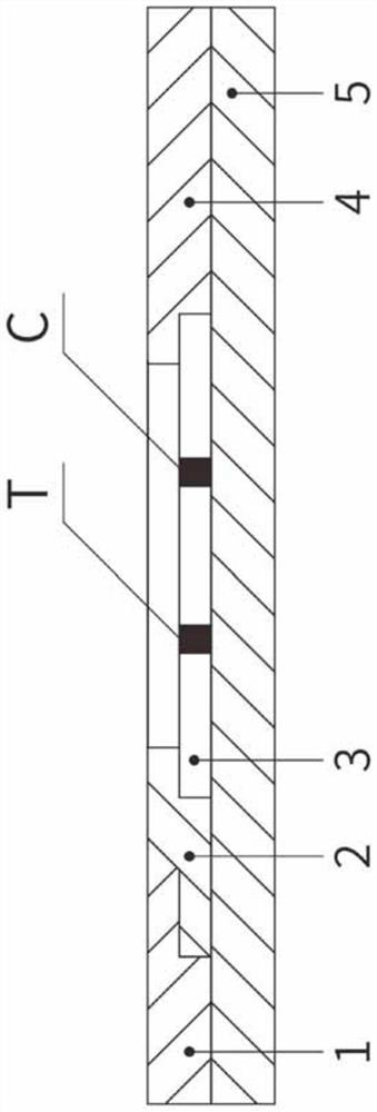
[0038] The sample pad 1, the immunofluorescent glass fiber membrane 2, the immunonitrocellulose membrane 3, and the absorbent pad 4 are respectively pasted on the plastic plate 5, and the two ends of the immunonitrocellulose membrane 3 are connected to the absorbent pad 4, the immunofluorescent glass fiber membrane, respectively. 2 overlapping, the other end of the immunofluorescence glass fiber membrane 2 is overlapped with the sample pad 1; a detection line T and a quality control line C are set on the immunonitrocellulose membrane 3, and a solid phase on the detection line T There is GDF15-antibody; the quality control line C is sprayed with goat anti-rabbit IgG polyclonal antibody;

[0039] Preparation:

[0040] (a), the preparation of immunofluorescent microspheres, take 100ul of microsphere suspension with a solid content of 1%, dilute 10 times with ultrapure water, that is, 1000ul, add it to the EP tube, take 40ul of N-hydroxysuccinyl Add the amine solution NHS to the ...

Embodiment 2

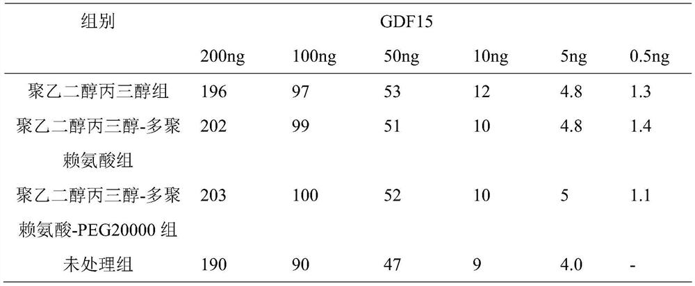
[0057] (b) Activated immunofluorescence microsphere cross-linked labeled antibody, take 1ml of activated microsphere suspension, disperse evenly by ultrasonic, then add antibody dropwise while stirring, after adding antibody, react for 75s, and then ultrasonically sonicate 30 seconds, then react for 1 hour, add BSA to block for 1 hour, centrifuge the blocked microspheres at a speed of 10000r / min for 15min, add immunofluorescence microsphere buffer to the centrifuged microspheres, and make The microspheres are uniformly dispersed and ready for use. The fluorescent buffer solution includes: a concentration of 20mM Tris-HCL solution, a sucrose concentration of 12%, a trehalose concentration of 3%, a BSA concentration of 0.7%, and a pH of 8.5;

[0058] Preparation of polyethylene glycol glycerin treatment solution: mixed with polyethylene glycol glycerin and polylysine (SIGMA, 150KD), wherein the concentration of polyethylene glycol glycerin is 0.5%, and polylysine The concentrati...

Embodiment 3

[0062] (b) Activated immunofluorescence microsphere cross-linked labeled antibody, take 1ml of activated microsphere suspension, disperse evenly by ultrasonic, then add antibody dropwise while stirring, after adding antibody, react for 120s, and then ultrasonically sonicate 40 seconds, then react for 1 hour, add BSA to block for 1 hour, centrifuge the blocked microspheres at a speed of 10000r / min for 15min, add the immunofluorescence microsphere buffer to the centrifuged microspheres, and make The microspheres are uniformly dispersed and ready for use. The fluorescent buffer solution includes: 20mM Tris-HCL solution, 20% sucrose concentration, 5% trehalose concentration, 1% BSA concentration, and a pH of 8.5;

[0063] Preparation of polyethylene glycol glycerin treatment solution: mixed with polyethylene glycol glycerin, polylysine (SIGMA, 150KD) and PEG2000, wherein the concentration of polyethylene glycol glycerin is 0.5%, poly The concentration of lysine is 0.5%, the concentr...


PUM


Abstract

The invention relates to a GDF15 rapid quantitative fluorescent microsphere detection device and a preparation method thereof, and belongs to the field of medical detection. The device is prepared byadhering a nitrocellulose membrane with a solid phase containing a GDF15 antibody and a goat-anti-mouse IgG polyclonal antibody, glass fiber adsorbed with a fluorescent microsphere labeled GDF15 antibody, a sample pad, absorbent paper and other auxiliary materials. The preparation method comprises the following steps: pretreating a nitrocellulose membrane by adopting a polyethylene glycol glyceroltreatment solution; combining a GDF15 antibody firstly with oleic acid modified zinc sulfide nanoparticles and then adsorbing to a nitrocellulose membrane; and preparing the proper buffer solution and the sample pad treatment solution. The reaction sensitivity is effectively improved on the basis of ensuring complete release of the immunofluorescence microsphere, and under the same threshold value, the dosage of the immunofluorescence microsphere can be reduced, the cost is saved, and the complexity of production operation is not increased. The test paper is high in sensitivity, strong in specificity, simple and convenient to operate, time-saving and strong in practicability.

Description

technical field [0001] The invention relates to the field of medical testing equipment, in particular to a GDF15 rapid quantitative fluorescent microsphere detection device and a preparation method thereof, which utilizes fluorescence immunochromatography technology and the principle of competition method to quantitatively detect the content of GDF15 in whole blood, serum, plasma and related liquid samples The device and the preparation method thereof can realize rapid detection of GDF15, and have high sensitivity and strong specificity. Background technique [0002] In the field of medicine, accurate early warning and assessment of abnormal conditions in the body have always been one of the main topics for medical workers to study and overcome; for example, what risks will happen to the human body under normal physiological conditions; Early warning, how to accurately assess disease outcome or development, etc.; therefore, it is very necessary to achieve the above goals thr...

Claims


Application Information

Patent Timeline
Patent Type & Authority Applications(China)
IPC IPC(8): G01N33/68G01N33/558G01N33/58G01N33/543
CPCG01N33/54346G01N33/558G01N33/582G01N33/74G01N2333/495
Inventor 杨小军李欣
Owner JILIN PROVINCE GERUISITE BIOTECHNOLOGY CO LTD
Why Eureka
Try Eureka