Magnetic-assisted built-in SERS microchip and preparation method thereof

A microchip and magnetic technology, applied in the field of biosensing, can solve the problems of many SERS substrate manufacturing steps, limited detection flux, and high operation requirements, and achieve the effect of high production cost, low cost, and high operation requirements

Active Publication Date: 2020-09-18
SOUTHEAST UNIV
View PDF5 Cites 1 Cited by
  • Summary
  • Abstract
  • Description
  • Claims
  • Application Information

AI Technical Summary

Problems solved by technology

The SERS substrate of the invention has many manufacturing steps (mercapto-functionalized grooves, mask covering, photoetching, in-situ gold deposition lamps), high cost, high operation requirements, and is manufactured one needle at a time. There is a batch-to-batch error in the SERS substrate; different response molecules are en

Method used


Image

  • Magnetic-assisted built-in SERS microchip and preparation method thereof
  • Magnetic-assisted built-in SERS microchip and preparation method thereof
  • Magnetic-assisted built-in SERS microchip and preparation method thereof

Examples


Embodiment 1

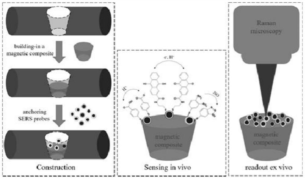
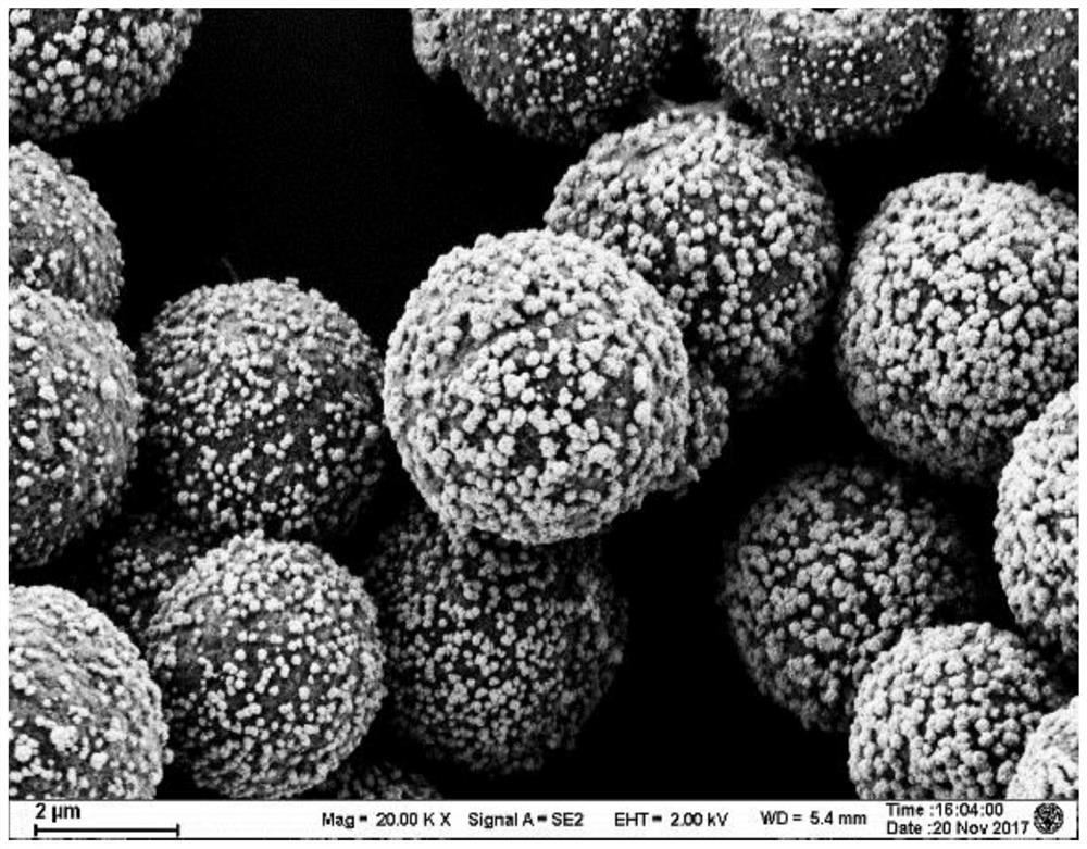
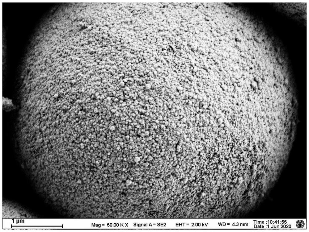
[0037] Example 1 Preparation of magnetic SERS active material—magnetic bead gold-shell composite

[0038] Synthesis of magnetic surface-enhanced Raman scattering (SERS) active materials: First, 2-3nm gold nanoparticles are adsorbed on magnetic beads with amino groups with a diameter of 5-6 microns to form composite particles, and the formed composite particles are magnetic surface-enhanced Raman scattering (SERS) active material precursor; hydrogen peroxide is used as a reducing agent to continuously reduce chloroauric acid under the catalysis of gold nanoparticles on the surface of the precursor and continuously deposit on its surface to form a complete magnetic bead Gold Shell Composite. The magnetic bead gold-shell complex was magnetically separated, the supernatant was discarded, and the precipitate was collected to obtain the magnetic bead gold-shell complex. figure 2 and image 3 The electron microscope image of the magnetic bead gold-shell composite is shown. The diame...

Embodiment 2

[0039] Example 2 Preparation of pH-responsive SERS probes

[0040] The magnetic SERS active material prepared in Example 1, the magnetic bead gold-shell composite, was immersed in a 1 mM ethanol solution of mercaptobenzoic acid for 30 minutes, and the mercaptobenzoic acid was bound to the surface of the Raman-enhanced nanomaterial to obtain a pH-responsive SERS probe.

Embodiment 3

[0041] Example 3 Redox State Responsive SERS Probe Preparation

[0042] First, 100mg of 2-carboxyanthraquinone, 80mg of dicyclohexylcarbodiimide and 115mg of N-hydroxysuccinimide were successively dissolved in 50mL of dimethyl sulfoxide and stirred at room temperature for three hours; then 22.5mg of cystamine was added Dihydrochloride, after stirring evenly, put it in a refrigerator at 4°C, and let it stand for 10 hours; then take out 0.1mL of the supernatant after the reaction and dilute it 100 times with ethanol, and the magnetic SERS active material prepared in Example 1—magnetic beads The gold-shell composite was immersed for 30 minutes to obtain a redox state responsive SERS probe.


PUM

PropertyMeasurementUnit
Diameteraaaaaaaaaa

Abstract

The invention discloses a magnetic-assisted built-in SERS microchip and a preparation method thereof. The preparation method comprises the following steps that: 1) forming a hole in an acupuncture needle, wherein the diameter of the hole is 10-80% of that of a needle body of the acupuncture needle; 2) filling part of the hole with a magnetic powder polymer compound, and forming a magnetic pit in an unfilled part after magnetizing the magnetic powder; and 3) additionally arranging a magnetic surface enhanced Raman scattering (SERS) probe into the magnetic pit. The magnetic-force-assisted built-in SERS microchip can be used for in-vivo sampling and in-vitro signal reading of various markers. The magnetic-assisted built-in SERS microchip is especially suitable for detection of biomarkers withshort half-life period, biomarkers which are easy to deteriorate (such as oxidation) when exposed to air, biomarkers which cannot be detected in vitro and the like. The magnetic-assisted built-in SERS microchip has the advantages of rapidness, simplicity, convenience, low detection limit, high selectivity, high sensitivity and the like in use.

Description

technical field [0001] The invention belongs to the technical field of biosensing, and in particular relates to a magnetic-assisted built-in SERS microchip and a preparation method thereof. Background technique [0002] Acupuncture needle-based detection technology is a minimally invasive in vivo or ex vivo detection technology. In 2002, Ren et al reported an electrochemical detection method based on acupuncture needles to construct calcium ion sensing needles. Since 2015, Zhang GJ et al. have successively reported the electrochemical detection method of dopamine, 5-hydroxytryptamine and NO sensor needles based on acupuncture needles. The biggest advantage of the detection technology based on acupuncture needles is that it can be minimally invasive in and out of the living body to achieve in-vivo detection or in-vivo sampling and ex-vivo detection. If multi-throughput detection can be realized, the electrochemical detection method will be an ideal in vivo detection method....

Claims


Application Information

Patent Timeline
IPC IPC(8): G01N21/65A61B10/00A61H39/08
CPCG01N21/658A61B10/00A61H39/086G01N2021/656
Inventor 董健陈一初李哲潘晨嫣
Owner SOUTHEAST UNIV
Why Eureka
Try Eureka