Multi-column for isolating exosomes and exosome isolation method
A separation method and technology of exosomes, applied in separation methods, solid adsorbent liquid separation, peptide preparation methods, etc., can solve problems such as long time, loss of exosomes, and in-depth analysis of lipoprotein elution. , to achieve the effect of improving purity and excellent separation efficiency
- Summary
- Abstract
- Description
- Claims
- Application Information
AI Technical Summary
Problems solved by technology
Method used
Image
Examples
Embodiment 1
[0064] Embodiment 1: the preparation of multicolumn
[0065] Sepharose (Sepharose) CL-6B was filled with 70% (v / v) in the lower part (diameter: 10mm, height: 50mm) of the column used for chromatography, and acryl-dextran gel (Sephacryl) 200 - HR was filled in its upper part at 30% (v / v), so that a multi-column was prepared so that the height ratio of the lower part and the upper part was 7:3. Here, the average surface charge values of Sepharose CL-6B and Sephadex 200-HR were -30 and -7, respectively.
Embodiment 2
[0066] Example 2: Confirmation of Exosome Separation Ability from Existing Column Conditions
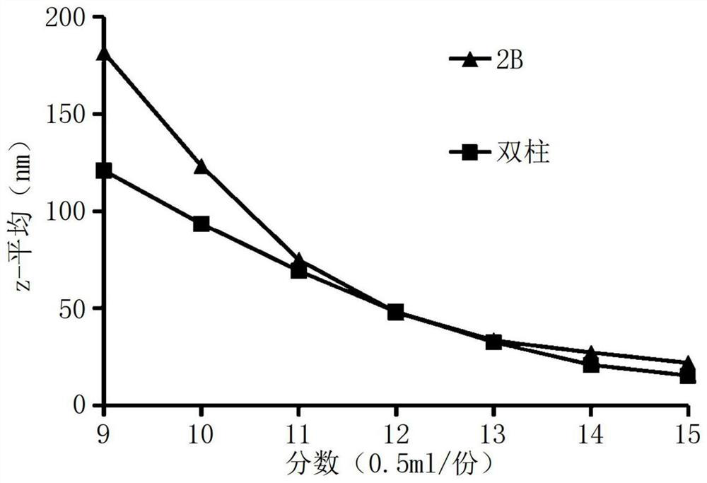
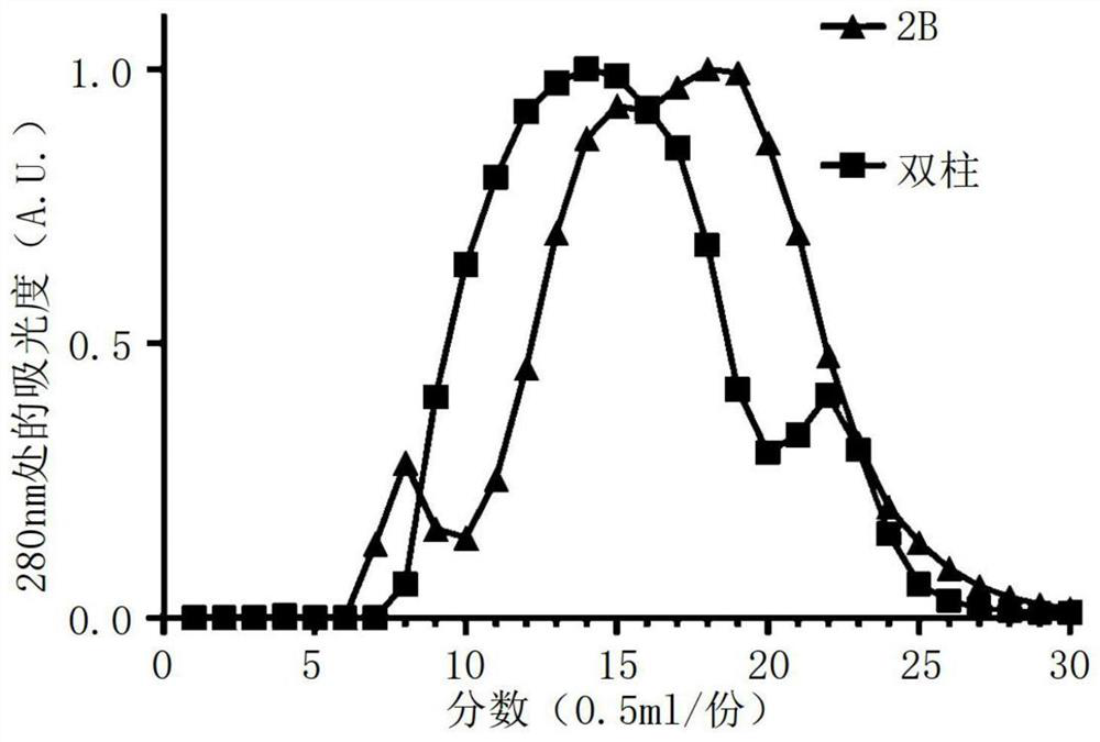
[0067] A column for chromatography packed with beads (diameter: 10 mm, height: 50 mm) was used, and as a sample, supernatant serum produced after centrifugation at 10000 rcf at 4° C. for 30 minutes was used. Size Exclusion Chromatography (SEC, Size Exclusion Chromatography) was performed on the samples in order to determine the separation efficiency of the material by existing single and multi-columns.
[0068] Specifically, as the beads packed in the column, Sepharose CL-2B was used as a single column (2B), and (Dual column) prepared in Example 1 was used as a multi-column. Beads filled with 300 ml PBS were washed before loading samples and the prepared samples were loaded just in time when the mobile phase buffer (except packed column beads) was exhausted. The volume of sample loaded on the column should not exceed 5% of the total volume of the packed beads, and 0.3 to 0.5 ml of...
Embodiment 3
[0070] Example 3: Comparison of the elution trends of exosomes and lipoproteins in serum
[0071] In order to compare the elution of exosomes, lipoproteins and water-soluble proteins in each segment in serum samples, the multi-column of Example 1 was used to fractionate by the method described in Example 2, and each Fractions were subjected to SDS-PAGE and Western blot. As the primary antibody, an anti-CD63 antibody (sc-15363 manufactured by Santa Cruz) as an exosome marker protein was diluted at 1:500, and an ApoB-100 antibody (sc-15363 manufactured by Santa Cruz) as a lipoprotein antibody was used. 25542) was diluted 1:1000, and then the diluted antibody was used to confirm its elution.
[0072] As a result, albumin (˜65 kDa) was detected as a water-soluble protein after the 13th segment ( FIG. 3 ), and lipoproteins appeared most prominently in the 9th segment and were then detected until the 11th segment. After a small amount of exosomes were detected in the 9th segment...
PUM
| Property | Measurement | Unit |
|---|---|---|
| porosity | aaaaa | aaaaa |
| size | aaaaa | aaaaa |
| diameter | aaaaa | aaaaa |
Abstract
Description
Claims
Application Information
 - R&D
- Intellectual Property
- Life Sciences
- Materials
- Tech Scout
- Unparalleled Data Quality
- Higher Quality Content
- 60% Fewer Hallucinations
Browse by: Latest US Patents, China's latest patents, Technical Efficacy Thesaurus, Application Domain, Technology Topic, Popular Technical Reports.
© 2025 PatSnap. All rights reserved.Legal|Privacy policy|Modern Slavery Act Transparency Statement|Sitemap|About US| Contact US: help@patsnap.com


