
NIR-II imaging probe and preparation method and application thereof

An imaging probe and probe technology, applied in the field of in vivo fluorescence imaging of cells, can solve the problems of inability to identify the boundary between tumor and healthy tissue, increase in recurrence and death, blurred edge of tumor site, etc., to reduce non-specific cell phagocytosis, The effect of prolonging blood circulation, high luminescence performance and photostability

Pending Publication Date: 2020-10-16
NANJING UNIV OF POSTS & TELECOMM
View PDF1 Cites 1 Cited by
  • Summary
  • Abstract
  • Description
  • Claims
  • Application Information

AI Technical Summary

Problems solved by technology

[0004] Ag now 2 Due to its excellent optical properties and biocompatibility in the second near-infrared region, S quantum dots are widely used as ideal fluorescent bioprobes in the second near-infrared region for fluorescence imaging, while the same dose of Ag 2 When S-labeled BMSCs were injected into mice with liver cancer, the NIR-II signal of the liver was obvious, but the edge of the tumor was blurred, and the boundary between the tumor and healthy tissue could not be identified, and the tumor could not be removed accurately and efficiently. Greatly increased chances of recurrence and death after surgery

Method used


Image

  • NIR-II imaging probe and preparation method and application thereof
  • NIR-II imaging probe and preparation method and application thereof
  • NIR-II imaging probe and preparation method and application thereof

Examples


Embodiment 1

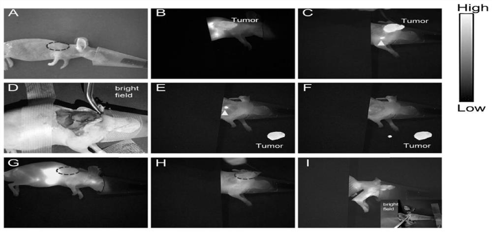
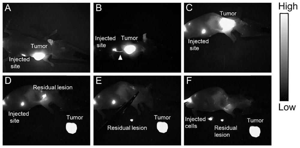
[0026] A NIR-II approach for tumor imaging and image-guided surgery.

[0027] S1. Put the mesenchymal stem cells in 25×25cm 2 culture flask with MSCM mesenchymal stem cell medium at 37 °C, CO 2 Cultivate under the condition of 5% concentration, until the cells grow to 80%-90%, about 5×10 6 , discard the culture medium, add 5ml of CP@mSiO with a concentration of 50μg / ml diluted with low-sugar DMEM incomplete medium 2 -PEG, incubated for 18-22h;

[0028] S2. Digest the cells with 2ml of 0.25% trypsin-EDTA solution for 1-2min, pay attention to observe the shape of the cells, and stop the digestion until the cells are completely detached; add 2-3ml of the medium to stop the digestion, mix the cell suspension Transfer to a 15ml centrifuge tube, centrifuge at 1000r for 5min, discard the supernatant waste liquid after centrifugation, add 500μl PBS to the cells, gently pipette, and resuspend the cells;

[0029] S3. Gently pipette the cell suspension evenly with a 1ml syringe, draw...

Embodiment 2

[0041] A NIR-II approach for tumor imaging and image-guided surgery.

[0042] S1. Put the mesenchymal stem cells in 25×25cm 2 culture flask with MSCM mesenchymal stem cell medium at 37 °C, CO 2 Cultivate under the condition of 5% concentration, until the cells grow to 80%-90%, about 5×10 6 , discard the culture medium, add 5ml of 50μg / ml diluted with low-sugar DMEM incomplete medium, and incubate for 18-22h;

[0043] S2. Digest the cells with 2ml of 0.25% trypsin-EDTA solution for 1-2min, pay attention to observe the shape of the cells, and stop the digestion until the cells are completely detached; add 2-3ml of medium to stop the digestion, mix well and suspend the cells Transfer the solution to a 15ml centrifuge tube, centrifuge at 1000r for 5min, discard the upper waste solution after centrifugation, add 500μl PBS to the cells, gently pipette, and resuspend the cells;

[0044] S3. Gently pipette the cell suspension evenly with a 1ml syringe, draw 200 μl of the cell suspe...


PUM


Abstract

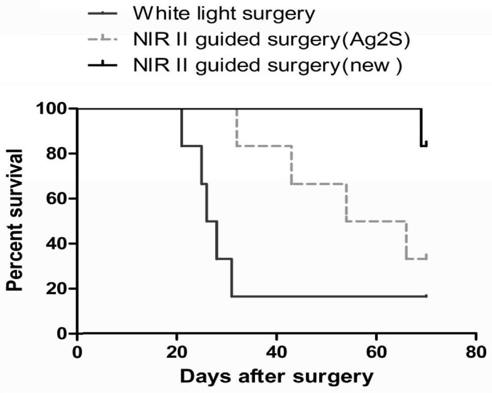
The invention discloses a NIR-II imaging probe and a preparation method and application thereof, and belongs to the technical field of in vivo bioluminescence imaging of cells. The invention comprisesthe following steps: S1, incubating bone marrow mesenchymal stem cells (BMSCs) of fluorescent probe CP@mSiO2(subscript)-PEG nanoparticles; S2, injecting fluorescent material-labeled BMSCs into HepG2tumor-bearing nude mice through tail vein injection and local subcutaneous injection; and S3, carrying out near-infrared-II (NIR-II) window imaging on the tumor, and then performing imaging-guided tumor resection. In the study, NIR-II is a new strategy for tumor imaging in vivo and image-guided tumor resection surgery, the NIR-II in vivo bioluminescence imaging technology is used to induce the fluorescent material-labeled BMSCs to enrich in the tumor site, so that the outline of the tumor can be seen clearly, thus improving the accuracy of surgical resection and reducing the tumor recurrence rate and the mortality rate.

Description

technical field [0001] The invention belongs to the technical field of living cell fluorescence imaging, and in particular relates to a NIR-II imaging probe and a preparation method and application thereof. [0002] technical background [0003] In the process of individual development, stem cells have the ability to self-replicate, and differentiate under certain conditions to form primitive cells of various types of cells. Mesenchymal stem cells have been widely used in the fields of stem cell transplantation, tissue engineering and immunotherapy, and tissue engineering repair in important organ damage / failure (liver failure, myocardial infarction, bone and cartilage defects, skin and soft tissue defects, lymphatic system function Obstacles, etc.) have excellent application prospects in reconstruction therapy; compared with visible light and near-infrared windows, the second near-infrared window (NIR-II) has higher tissue penetration, temporal resolution, and spatial The a...

Claims


Application Information

Patent Timeline
Patent Type & Authority Applications(China)
IPC IPC(8): A61K49/00C09K11/06
CPCA61K49/0021A61K49/0054C09K11/06C09K2211/1051C09K2211/1092
Inventor 张磊白洁琼寿康全陆峰范曲立黄维
Owner NANJING UNIV OF POSTS & TELECOMM
Learn More