
Protoporphyrin liposome for nuclear delivery of chemotherapeutic drugs and preparation method and application of protoporphyrin liposome

A chemotherapeutic drug and porphyrin lipid technology, which is applied in the field of protoporphyrin liposomes for chemotherapeutic drug cell nucleus delivery and its preparation, can solve the problem of changing the subcellular localization of chemotherapeutic drugs, reducing the anticancer effect of doxorubicin, and difficulty in exerting chemotherapeutic effects. To achieve the effect of nuclear drug delivery, improve tumor treatment effect, good stability and drug loading effect

Active Publication Date: 2020-11-13
SOUTHEAST UNIV
View PDF5 Cites 0 Cited by
  • Summary
  • Abstract
  • Description
  • Claims
  • Application Information

AI Technical Summary

Problems solved by technology

However, the final target of most common chemotherapeutic drugs (such as doxorubicin, cisplatin, and camptothecin) is the nucleus, and the encapsulation of nanocarriers often changes the subcellular localization of chemotherapeutic drugs, allowing them to enter the nucleus. greatly reduced efficiency
The process of drug uptake by cells is very complicated. Many nanomedicines are inevitably captured by lysosomes after entering the cells and it is difficult to escape from lysosomes. Moreover, there are nuclear pore complexes on the surface of the nucleus, which usually only allow small molecular weight substances to enter and exit freely. Nuclei, while substances with larger molecular weights have difficulty passing through the nuclear pore complex
Therefore, it is difficult for many nanocarriers to release the loaded small molecule drugs in time after delivering the drugs to the tumor area, making it difficult for the drugs to enter the nucleus and play a role
For example, liposomal doxorubicin (DOXIL), which is currently used clinically, has reduced cardiotoxicity and higher tumor enrichment efficiency than doxorubicin, but after it reaches the tumor area, the doxil loaded in it enters the tumor cell nucleus. However, the efficiency is very low, making it difficult for most of the doxorubicin molecules to play a chemotherapeutic role, thus greatly reducing the anticancer effect of doxorubicin

Method used


Image

  • Protoporphyrin liposome for nuclear delivery of chemotherapeutic drugs and preparation method and application of protoporphyrin liposome
  • Protoporphyrin liposome for nuclear delivery of chemotherapeutic drugs and preparation method and application of protoporphyrin liposome
  • Protoporphyrin liposome for nuclear delivery of chemotherapeutic drugs and preparation method and application of protoporphyrin liposome

Examples


Embodiment 1

[0043] Step 1: 13.07 mg of hydrogenated soybean lecithin, 3.70 mg of distearoylphosphatidylethanolamine-polyethylene glycol 2000, 3.23 mg of cholesterol and 0.74 mg of protoporphyrin were co-dissolved in 1 mL of a mixed organic solvent of chloroform and methanol (chloroform: Methanol=65:35, volume ratio), and mix well;

[0044] Step 2: The mixed solution in step 1 is blown dry with nitrogen and vacuum-dried for 12 hours to form a film;

[0045] Step 3: Hydrate the film formed in step 2 with 5 mL of phosphate buffer, and perform probe ultrasound at 60°C (ultrasonic power: 163W, total ultrasonic time: 40s (ultrasonic 4s, stop 2s, repeat 10 times) ) to form a protoporphyrin liposome dispersion.

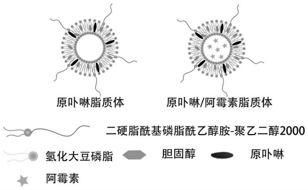
[0046] The structure of the protoporphyrin liposome prepared in this embodiment is shown in figure 1 . The liposome forms a vesicle structure as a whole, and the interior is a water cavity, and both phospholipid molecules (HSPC) and DSPE-PEG can provide hydrophobic tails.

Embodiment 2

[0048] The stability evaluation of the protoporphyrin liposome prepared by embodiment 1:

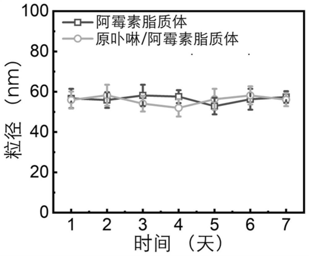
[0049] The protoporphyrin liposome dispersion liquid obtained in Example 1 and the liposome dispersion liquid not containing protoporphyrin (protoporphyrin was not added in the preparation process of Example 1) were respectively in volume ratio 1:9 and containing 10% Fetal bovine serum and phosphate buffer were mixed and placed at 37°C. The particle size of the two liposomes was monitored over a week. The particle size of liposomes was measured by a potentiometric-particle size analyzer.

[0050] see results figure 2 , the particle size of the two liposomes had no significant change within a week, indicating that both the protoporphyrin liposome and the blank liposome had good stability.

Embodiment 3

[0052] The encapsulation of chemotherapeutic drug doxorubicin by the protoporphyrin liposome prepared in embodiment 1:

[0053] Step 1: 13.07 mg of hydrogenated soybean lecithin, 3.70 mg of distearoylphosphatidylethanolamine-polyethylene glycol 2000, 3.23 mg of cholesterol and 0.74 mg of protoporphyrin were co-dissolved in 1 mL of a mixed organic solvent of chloroform and methanol (chloroform: Methanol=65:35, volume ratio), and mix well;

[0054] Step 2: The mixed solution in step 1 is blown dry with nitrogen and vacuum dried for 12 hours to form a film;

[0055] Step 3: Dissolve 5 mg of doxorubicin hydrochloride in 5 mL of phosphate buffer to obtain a 1 mg / mL doxorubicin solution;

[0056] Step 4: Mix the film formed in step 2 with all the doxorubicin solution in step 3, and perform probe ultrasound at 60°C (ultrasonic power: 163W, total ultrasonic time: 40s (ultrasonic 4s, stop 2s, repeat 10 times)) to form a liposome dispersion.

[0057] Step 5: The liposome dispersion f...


PUM


Abstract

The invention discloses protoporphyrin lipidosome for nuclear delivery of chemotherapeutic drugs and a preparation method and application of the protoporphyrin lipidosome. The protoporphyrin lipidosome is mainly prepared from hydrogenated soybean lecithin, distearoyl phosphatidylethanolamine-polyethylene glycol 2000, cholesterol and protoporphyrin. The liposome can be prepared by a film hydrationmethod and further ultrasonic or extrusion treatment, and can wrap various chemotherapeutic drugs to form protoporphyrin liposome loaded chemotherapeutic drugs so as to realize in-vivo long circulation and tumor delivery of the drugs. Moreover, compared with the traditional liposome, the liposome can increase the cell nucleus distribution of the chemotherapeutic drug, realizes more efficient cellnucleus drug delivery and cell killing, has good safety and good universality, and is expected to become a universal chemotherapeutic drug cell nucleus delivery carrier.

Description

technical field [0001] The invention belongs to biotechnology, and in particular relates to a protoporphyrin liposome used for delivering chemotherapeutic drugs to cell nucleus and its preparation method and application. Background technique [0002] Chemotherapy is currently one of the main means of clinical treatment of cancer. However, the poor targeting of chemotherapeutic drugs and low tumor enrichment efficiency lead to limited therapeutic effect and high systemic toxicity. The delivery of chemotherapy drugs through nanocarriers can effectively prolong the circulation time of chemotherapy drugs in vivo and improve the tumor accumulation efficiency of drugs. However, the final target of most common chemotherapeutic drugs (such as doxorubicin, cisplatin, and camptothecin) is the nucleus, and the encapsulation of nanocarriers often changes the subcellular localization of chemotherapeutic drugs, allowing them to enter the nucleus. Efficiency is greatly reduced. The proce...

Claims


Application Information

Patent Timeline
Patent Type & Authority Applications(China)
IPC IPC(8): A61K47/69A61K47/54A61K45/00A61K9/127A61P35/00A61K31/704A61K33/243A61K31/555A61K31/4745A61K31/337
CPCA61K47/6911A61K45/00A61P35/00A61K47/546A61K9/1277A61K31/704A61K33/243A61K31/555A61K31/4745A61K31/337Y02A50/30
Inventor 吴富根祝雅璇贾浩然段秋怡
Owner SOUTHEAST UNIV
Learn More