An automatic separation and enrichment device and method for tumor cells
A technology for separating and enriching tumor cells, which is applied in the field of automated separation and enrichment devices, and can solve the problems of clinical diagnosis deviation, low efficiency of separating whole blood, and cell damage.
- Summary
- Abstract
- Description
- Claims
- Application Information
AI Technical Summary
Problems solved by technology
Method used
Image

Examples
Embodiment 1
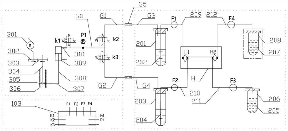
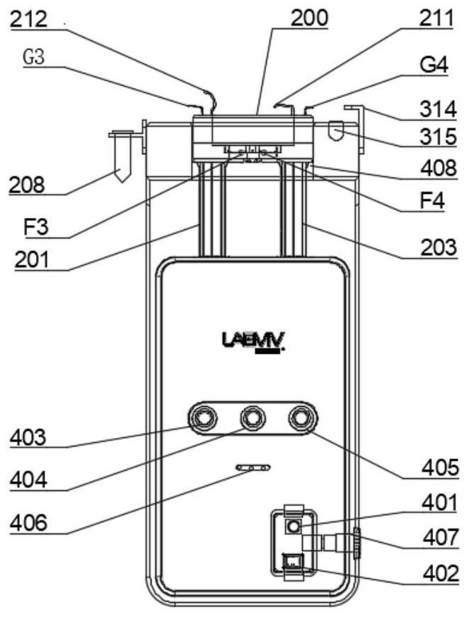
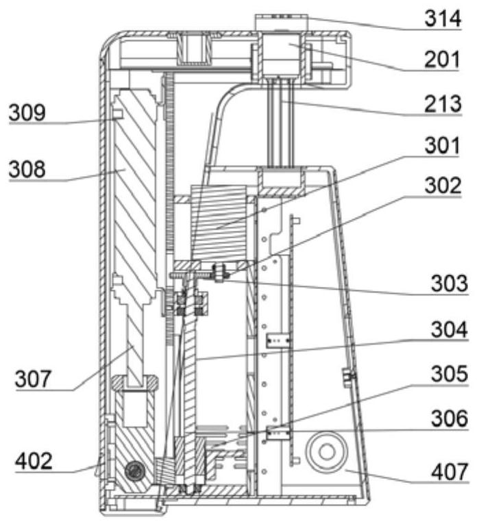
[0060] An operation method of an automated microfluidic system for enrichment of circulating tumor cells, the method adopts the method attached to the present invention Figure 1-4 equipment, the specific operations are as follows:
[0061] S01: Remove the sample tube and the buffer tube in the device respectively, add 2.3mL of ten-fold concentration of cell preservation solution to the buffer tube, then add deionized water to the buffer solution to the set scale, and mix well;
[0062] S02: Put the sample or blood collection tube to be tested into the sample tube, and add the mixed cell preservation solution to the sample tube to 5mL;
[0063] S03: Put the buffer tube and sample tube into the corresponding position of the device, put a 50mL centrifuge tube in the waste tube rack as the waste tube, and then put a 1.5mL or 2mL enrichment tube in the enrichment tube slot;
[0064] S04: Press the start button, wait for the sample to be loaded, and after the sample in the sample ...
Embodiment 2
[0067] Adopt the present invention to attach Figure 1-4 The equipment in the method separates different types of cell lines, and recovers the separated cell lines and calculates the recovery efficiency of the cell lines. The specific method is as follows:
[0068] 1) Take the non-small cell lung cancer cell line H1299 cultured in T25, discard the medium, add 5mL PBS to wash twice, add 1mL trypsin to make the trypsin evenly cover the cell surface, discard the excess trypsin, and incubate at 37 degrees Celsius for 3 Minutes, add medium containing 10% FBS to terminate the digestion reaction, mix the cells evenly with a 1mL pipette, and confirm under the microscope that all cells are dispersed into single cells. And take 100 μL of cells, use a cell counter for counting.
[0069] 2) According to the concentration calculated by the cell counter, use the cell preservation solution to dilute H1299. The dilution concentration is 100 H1299 cells in the 5mL dilution solution. Add the dil...
Embodiment 3
[0077] Adopt the present invention to attach Figure 1-4 The device in the method separates the cells and detects the activity of the separated cells, the specific method is as follows:
[0078] 1) Take the lung cancer cell line A549 cultured in T25, discard the medium, add 5mL PBS to wash twice, add 1mL trypsin to make the trypsin evenly cover the cell surface, discard the excess trypsin, incubate at 37 degrees Celsius for 3 minutes, add The medium containing 10% FBS stopped the digestion reaction, mixed the cells evenly with a 1mL pipette, and confirmed under the microscope that all the cells were dispersed into single cells. And take 100 μL of cells, add trypan blue according to 1:1, use a cell counter to count and calculate the cell viability; the measured initial cell viability is 96.81%.
[0079] 2) Adjust the cell concentration to 1×10⁶ cells / mL, and add the diluted cells to the attached Figure 1-3 in the sample tube of the device and use the device for separation. ...
PUM
 Abstract
Description
Claims
Application Information
 - R&D
- Intellectual Property
- Life Sciences
- Materials
- Tech Scout
- Unparalleled Data Quality
- Higher Quality Content
- 60% Fewer Hallucinations
Browse by: Latest US Patents, China's latest patents, Technical Efficacy Thesaurus, Application Domain, Technology Topic, Popular Technical Reports.
© 2025 PatSnap. All rights reserved.Legal|Privacy policy|Modern Slavery Act Transparency Statement|Sitemap|About US| Contact US: help@patsnap.com